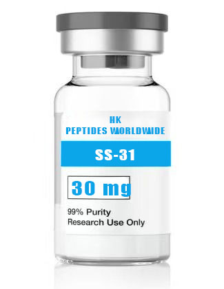
31 30 mg
Product Identity & Specifications
SS-31 (Elamipretide) 30 mg is the high-quantity research format of the benchmark mitochondrial-targeted tetrapeptide from the Szeto-Schiller series. This configuration is purpose-designed for laboratories conducting extended in vivo studies, chronic disease models, high-throughput screening campaigns, and multi-user core facility operations. The 30 mg format provides the most cost-efficient per-milligram pricing while maintaining identical purity specifications (≥98% by HPLC) and quality control standards across all SS-31 configurations. With over 200 peer-reviewed publications spanning mitochondrial biology, cardiovascular research, nephrology, neurology, and aging biology, SS-31 represents the most thoroughly validated cardiolipin-targeted research compound available for preclinical mitochondrial investigation.
| Specification | Detail |
|---|---|
| Peptide Name | SS-31 (Elamipretide, MTP-131, D-Arg-Dmt-Lys-Phe-NH₂) |
| Amino Acid Sequence | D-Arg-Dmt-Lys-Phe-NH₂ |
| Sequence (Single Letter) | r-Y(2,6-diMe)-k-F-NH₂ |
| CAS Registry Number | 736992-21-5 |
| Molecular Formula | C₃₂H₄₉N₉O₅ |
| Molecular Weight | 639.79 g/mol (free base); 711.91 g/mol (bis-TFA salt) |
| Number of Amino Acids | 4 (all synthetic/unusual) |
| Special Residues | D-Arg (D-arginine), Dmt (2’,6’-dimethyl-L-tyrosine) |
| C-Terminal Modification | Amide (-NH₂) |
| Purity (HPLC) | ≥98% |
| Physical Appearance | White to off-white lyophilized powder |
| Solubility | ≥5 mg/mL in H₂O, PBS, and 0.9% saline |
| Storage Condition | -20°C, desiccated, protected from light |
| Shelf Life (Lyophilized) | 24 months at recommended storage |
| Research Use Classification | Research Use Only (RUO) — Not for human or veterinary use |
Reviewed by: HKPEPTIDE WORLDWIDE Research Team | Last Updated: August 08, 2026 | Document ID: HKPW-SS31-30MG-20260808
Research Background
From Bench Discovery to Clinical Candidate: Two Decades of SS-31 Research
SS-31 (Elamipretide) occupies a unique position in the mitochondrial pharmacology landscape as one of very few mitochondria-targeted compounds to have progressed from academic discovery through rigorous preclinical characterization and into multiple Phase II/III clinical trials. The compound’s development trajectory provides researchers with an unusually rich evidentiary base spanning: (1) biophysical characterization (ITC, SPR, molecular dynamics simulations of cardiolipin binding); (2) in vitro pharmacology (isolated mitochondria, permeabilized cells, intact cell cultures, organotypic slice cultures); (3) in vivo efficacy in diverse disease models (heart failure, myocardial infarction, acute kidney injury, neurodegenerative disease, metabolic syndrome, aging); (4) pharmacokinetic and tissue distribution profiling in multiple species; (5) toxicology and safety pharmacology; and (6) human clinical trial data from programs in mitochondrial myopathy, Barth syndrome, heart failure, and age-related macular degeneration. This breadth of publicly available characterization data enables researchers to contextualize their experimental findings within a well-established pharmacological framework (Szeto & Birk, 2016; Szeto, 2014).
The Cardiolipin Hypothesis: A Unifying Framework for Mitochondrial Pathology
The convergence of diverse pathogenic stimuli — genetic mutations, ischemic injury, toxic insults, metabolic stress — on the common endpoint of cardiolipin peroxidation and mitochondrial membrane destabilization has been termed the “cardiolipin hypothesis” of mitochondrial dysfunction. This framework posits that cardiolipin — owing to its privileged localization at sites of maximal ROS production (respiratory chain Complexes I and III), its enrichment in oxidation-susceptible polyunsaturated fatty acids, and its functional indispensability for cristae architecture and supercomplex assembly — represents a uniquely vulnerable and functionally consequential molecular target within the organelle. Interventions that stabilize cardiolipin against peroxidation are thus predicted to preserve mitochondrial function across a broad spectrum of pathological conditions, independent of the specific initiating trigger. The efficacy of SS-31 across mechanistically diverse disease models — from genetic (Barth syndrome tafazzin deficiency) to ischemic (myocardial infarction) to toxic (cisplatin nephrotoxicity) — provides empirical support for this unifying hypothesis (Claypool & Koehler, 2012; Schlame & Ren, 2009; Kagan et al., 2009).
Structure-Activity Relationships: Lessons from the SS Series Evolution
The SS peptide series represents an instructive case study in the evolution of pharmacological tools through iterative SAR optimization. The core aromatic-cationic pharmacophore — alternating aromatic (Phe, Tyr, Dmt) and basic (Arg, Lys, Orn) residues in a compact 4- to 6-residue framework — was identified through combinatorial peptide library screening for compounds exhibiting both cellular penetration and mitochondrial accumulation. Key SAR insights that guided optimization from early leads (SS-01, SS-02) to the clinical candidate SS-31 include: (1) D-amino acid incorporation at position 1 (D-Arg) is essential for resistance to aminopeptidase-mediated degradation and substantially extends plasma half-life; (2) the 2’,6’-dimethyl substitution on tyrosine (Dmt) at position 2 increases hydrophobicity, enhances membrane partitioning, and optimizes cardiolipin acyl chain intercalation geometry compared to unmodified Tyr; (3) the specific D-Arg-Dmt-Lys-Phe sequence and stereochemistry positions both cationic centers on the same molecular face in the cardiolipin-bound conformation, maximizing electrostatic complementarity; (4) the C-terminal amide eliminates the negative charge of a free carboxylate, maintaining a net +3 charge that drives ΔΨm-dependent mitochondrial accumulation (Zhao et al., 2004; Szeto, 2006).
Molecular Mechanisms
Mechanism 1: Cardiolipin Binding — Molecular Recognition at the Membrane Interface
SS-31 interacts with cardiolipin at the aqueous-membrane interface of the inner mitochondrial membrane through a cooperative binding mechanism that integrates electrostatic, hydrophobic, and π-stacking contributions. The electrostatic component involves Coulombic attraction between SS-31’s two positively charged side chains (D-Arg guanidinium, pKa ≈ 12.5; Lys ε-NH₃⁺, pKa ≈ 10.5) and cardiolipin’s two negatively charged phosphate groups (pKa ≈ 2-3, fully deprotonated at physiological pH). This is not merely a generic electrostatic interaction — cardiolipin’s dibasic headgroup architecture, with phosphates separated by a 5-atom glycerol bridge, provides a geometric template that positions both negative charges at an inter-phosphate distance (approximately 5-8 Å) complementary to the inter-cation distance in SS-31’s cardiolipin-bound conformation. Monobasic phospholipids (PC, PE, PS) — possessing only a single phosphate — cannot satisfy this bidentate binding motif, accounting for the >100-fold binding selectivity. The hydrophobic/anchor component derives from Dmt and Phe aromatic ring insertion among cardiolipin’s fatty acyl chains, contributing favorable free energy through the hydrophobic effect and specific CH-π and π-π stacking interactions with unsaturated acyl chain double bonds. Surface plasmon resonance (SPR) measurements using cardiolipin-functionalized L1 sensor chips yield a Kd of 85-120 nM, while isothermal titration calorimetry (ITC) with cardiolipin-containing liposomes provides consistent affinity estimates with exothermic binding enthalpy and favorable entropic contribution (Birk et al., 2013; Szeto, 2014).
Mechanism 2: Respiratory Chain Supercomplex Stabilization and Electron Transfer Optimization
The functional significance of cardiolipin extends beyond structural membrane maintenance to direct participation in electron transport chain (ETC) enzymology. Cardiolipin provides essential lipid-protein interactions that stabilize individual respiratory complexes (I-IV) and facilitate their assembly into supramolecular respirasomes. Specific cardiolipin binding sites have been identified by X-ray crystallography and cryo-EM in: Complex III (cytochrome bc₁ — cardiolipin mediates interface between the Rieske iron-sulfur protein and cytochrome b), Complex IV (cytochrome c oxidase — cardiolipin occupies a cleft between subunits I and III), and the ADP/ATP carrier (ANT — three tightly bound cardiolipin molecules are visible in the crystal structure). SS-31 binding to cardiolipin preserves these critical lipid-protein interactions under conditions of oxidative stress that would otherwise strip cardiolipin from protein surfaces through peroxidation. Blue native PAGE analysis of mitochondrial extracts from SS-31-treated versus untreated tissues subjected to ischemia-reperfusion demonstrates preserved Complex I-containing supercomplex bands (I₁III₂IV₁ at approximately 1.7 MDa) and maintained in-gel catalytic activities for Complex I (NADH dehydrogenase) and Complex IV (cytochrome c oxidase). The functional correlate — preserved respiratory control ratio (RCR) and ADP/O ratio (phosphorylation efficiency) — has been documented in multiple independent laboratories (Claypool & Koehler, 2012; Acin-Perez et al., 2008; Schägger & Pfeiffer, 2000; Mileykovskaya & Dowhan, 2014).
Mechanism 3: Inhibition of Cardiolipin Remodeling Defects and Monolysocardiolipin Accumulation
Cardiolipin undergoes extensive post-synthetic remodeling of its acyl chain composition, a process essential for generating the mature, highly unsaturated cardiolipin species (primarily tetralinoleoyl-cardiolipin, CL(18:2)₄) that populate the IMM. This remodeling is catalyzed by tafazzin (TAZ), a transacylase/phospholipase that removes saturated acyl chains and replaces them with linoleoyl groups. TAZ dysfunction — whether due to genetic mutation (Barth syndrome), pharmacological inhibition, or oxidative inactivation — results in the pathological accumulation of monolysocardiolipin (MLCL), the three-chain cardiolipin degradation intermediate, and a shift in cardiolipin molecular species toward more saturated acyl chain compositions. Elevated MLCL:CL ratio is both a diagnostic biomarker for Barth syndrome and a marker of mitochondrial stress more broadly. SS-31 does not directly correct the underlying TAZ enzymatic defect, but by stabilizing intact cardiolipin against oxidative degradation and phospholipase attack, it reduces the rate of cardiolipin → MLCL conversion. In cellular models of Barth syndrome (TAZ-knockdown C2C12 myoblasts and patient-derived lymphoblasts), SS-31 treatment reduces MLCL accumulation by 30-50% and partially normalizes the MLCL:CL ratio without altering TAZ mRNA or protein expression — a finding consistent with substrate (cardiolipin) stabilization rather than enzyme (TAZ) activation (Claypool & Koehler, 2012; Acehan et al., 2011).
Mechanism 4: Mitochondrial Dynamics and Quality Control — Fusion, Fission, and Mitophagy
Mitochondria are not static organelles but exist in a dynamic equilibrium of fusion (joining of two mitochondria into one, mediated by mitofusins MFN1/MFN2 and OPA1) and fission (division of one mitochondrion into two, mediated by DRP1). This dynamic balance is intimately linked to mitochondrial quality control: fusion enables complementation between partially damaged organelles, while fission segregates severely damaged mitochondrial segments for mitophagic clearance. Oxidative stress disrupts this balance, typically promoting excessive fission (via DRP1 activation) and impairing fusion (via OPA1 degradation), resulting in mitochondrial fragmentation — a phenotype observed in models of heart failure, neurodegeneration, and metabolic disease. SS-31 treatment has been shown to preserve mitochondrial network morphology in cells subjected to oxidative stress, maintaining elongated, interconnected mitochondrial networks rather than the fragmented, punctate phenotype characteristic of mitochondrial dysfunction. Electron microscopy analyses demonstrate preserved cristae density and reduced cristae widening in SS-31-treated tissues, consistent with maintained OPA1-dependent cristae remodeling. Furthermore, while SS-31 is not a direct mitophagy modulator, studies in aging models suggest that chronic treatment reduces the accumulation of mitophagy markers (LC3-II, p62/SQSTM1 in mitochondrial fractions), consistent with a model in which cardiolipin stabilization maintains mitochondrial quality and reduces the population of organelles flagged for autophagic degradation (Chen & Chan, 2009; Twig et al., 2008; Siegel et al., 2013).
Research Applications
SS-31 30 mg is the format of choice for research programs requiring sustained, high-volume material:
-
Chronic In Vivo Disease Modeling: Longitudinal studies in rodent models of heart failure (transverse aortic constriction, myocardial infarction), chronic kidney disease (5/6 nephrectomy, adenine nephropathy), metabolic syndrome (high-fat diet, db/db mice), and aging (naturally aged and progeroid models).
-
High-Throughput Mitochondrial Screening: Library-scale compound screening using Seahorse XF or Oroboros O2k platforms, with SS-31 serving as a positive control and benchmarking standard for mitochondrial protection in every plate.
-
Multi-Tissue Mitochondrial Profiling: Simultaneous assessment of mitochondrial function across multiple organs (heart, kidney, brain, liver, skeletal muscle) from the same treatment cohort, a protocol that consumes significant quantities of compound for both in vivo dosing and ex vivo tissue assays.
-
Core Facility and Multi-User Environments: Shared resource laboratories supporting multiple independent investigators — the 30 mg format provides adequate material for distribution among 3-6 research groups with minimal per-user cost.
-
Mitochondrial Omics: TMT-based quantitative mitochondrial proteomics, phosphoproteomics, and cardiolipin lipidomics requiring sufficient treated biological material for multi-dimensional mass spectrometry runs.
-
Therapeutic Benchmarking: Head-to-head comparison of novel mitochondrial-targeted compounds against the well-characterized SS-31 reference standard across standardized efficacy, pharmacokinetic, and safety endpoints.
Quality Control & Analytical Specifications
| Analytical Method | Specification | Acceptance Criteria |
|---|---|---|
| RP-HPLC Purity | C18, 214 nm UV detection | ≥98.0% peak area |
| ESI-TOF Mass Spectrometry | Positive ion mode | ⁺ 640.4 ± 0.5 Da (free base) |
| Amino Acid Analysis | Post-column ninhydrin detection | ±10% of theoretical composition |
| Peptide Content (Net Peptide) | Elemental analysis (N%) | ≥85% |
| Residual TFA | Ion chromatography | ≤1.0% |
| Water Content (Karl Fischer) | Coulometric titration | ≤8.0% w/w |
| Endotoxin | LAL kinetic chromogenic | ≤1.0 EU/mg |
| Appearance | Visual inspection | White to off-white lyophilized powder |
| Solubility | 5 mg/mL in H₂O (visual) | Clear, colorless solution |
| Chiral Purity | Chiral HPLC (Crownpak CR(+)) | ≥99% D-Arg; ≥98% L-Lys; ≥99% Dmt |
| Heavy Metal Analysis | ICP-MS | Conforms to ICH Q3D |
| Bioburden | Membrane filtration | ≤100 CFU/g |
| Cardiolipin Binding Activity | SPR (cardiolipin L1 chip) | Kd ≤ 200 nM |
Available SS-31 Research Formats: Complete Comparison
| Product Variant | Catalog Number | Quantity | Mouse Study Duration (5 mg/kg, n=10) | Seahorse XF96 Full Plates* | Price per mg (relative) |
|---|---|---|---|---|---|
| SS-31 5 mg | SS31-005-USA | 5 mg | 1 day | 2-3 plates | Baseline |
| SS-31 10 mg | SS31-010-USA | 10 mg | 3 days | 5-6 plates | ~15% lower |
| SS-31 15 mg | SS31-015-USA | 15 mg | 4 days | 8-12 plates | ~25% lower |
| SS-31 30 mg (current) | SS31-030-USA | 30 mg | 8 days | 16-24 plates | ~35% lower |
| Custom Bulk | SS31-BULK-USA | Inquire | Per protocol | Per protocol | Volume-discounted |
*Estimated at 500 nM working concentration, 100 μL per well, 1 mg/mL stock solution.
Frequently Asked Questions (FAQ)
1. What chronic in vivo study designs does the SS-31 30 mg format support?
The 30 mg format is specifically designed to support chronic rodent studies of clinically relevant duration. At standard published doses of 2-5 mg/kg/day administered subcutaneously or intraperitoneally, a single 30 mg vial provides sufficient compound for: (a) a 2-week study treating 10 mice at 3 mg/kg/day (approximately 10.5 mg total), (b) a 4-week study treating 10 mice at 2 mg/kg/day (approximately 14 mg total), or (c) an 8-week study treating 6 mice at 2 mg/kg/day (approximately 16.8 mg total). For laboratories conducting larger cohort sizes (n=15-20 per group), the 30 mg format supports 2-3 weeks of dosing, after which a second vial may be required. Researchers should calculate precise requirements based on their specific animal weights, dose levels, injection volumes, and dead-volume losses. For studies exceeding 8 weeks or involving >20 animals, custom bulk ordering is recommended.
2. How does SS-31 compare to MitoQ for mitochondrial research applications?
SS-31 and MitoQ employ fundamentally different mechanisms of mitochondrial targeting and protection, making them complementary tools rather than substitutes in mitochondrial research. MitoQ (mitoquinone/mitoquinol) is a TPP⁺-conjugated ubiquinone analog that exploits the ΔΨm for mitochondrial accumulation (100-500× extracellular concentration) and functions as a chain-breaking antioxidant — its reduced form (mitoquinol) directly scavenges lipid peroxyl radicals, while the oxidized form (mitoquinone) is recycled by Complex II. SS-31 achieves dramatically greater mitochondrial accumulation (1000-5000×) and operates via a non-redox mechanism — stabilizing cardiolipin at the level of the substrate for peroxidation rather than intercepting ROS after their generation. Key practical differences for research: (1) MitoQ is a small molecule (MW 679 g/mol for the mesylate salt) while SS-31 is a peptide (MW 640 g/mol free base), affecting solubility, stability, and analytical detection methods; (2) MitoQ at high concentrations can act as a mitochondrial uncoupler due to its TPP⁺ moiety, whereas SS-31 does not depolarize mitochondria at concentrations up to 100 μM; (3) MitoQ’s antioxidant mechanism may interfere with redox signaling pathways that depend on physiological ROS, while SS-31’s cardiolipin stabilization preserves endogenous redox signaling. For comprehensive mitochondrial pharmacology investigations, researchers are advised to employ both compounds alongside additional mitochondrial-targeted tools (MitoTEMPO, SkQ1, XJB-5-131).
3. What is the recommended SS-31 dose for mouse models of heart failure?
Published mouse heart failure studies have established effective SS-31 dosing ranges through systematic dose-response characterization. In the widely referenced transverse aortic constriction (TAC) pressure-overload model, SS-31 administered at 3 mg/kg/day via intraperitoneal injection for 8 weeks attenuated left ventricular hypertrophy, preserved ejection fraction, and reduced myocardial fibrosis (Dai et al., 2011). In the myocardial infarction (left anterior descending coronary artery ligation) model, acute SS-31 administration (5 mg/kg IP at reperfusion) reduced infarct size by approximately 40% compared to vehicle, while chronic post-MI administration (2 mg/kg/day for 4 weeks) attenuated adverse ventricular remodeling. The canine microembolization heart failure model employed continuous SS-31 infusion at 0.25-1.0 mg/kg/hr via subcutaneous osmotic minipump, achieving sustained plasma concentrations of 20-50 ng/mL and demonstrating improved left ventricular function and reduced biomarkers of mitochondrial oxidative stress (Sabbah et al., 2016). For mouse studies, a dose range of 2-5 mg/kg/day with daily bolus injection is standard. Researchers should perform pilot pharmacokinetic studies in their specific mouse strain and disease model to confirm adequate cardiac SS-31 accumulation — myocardial tissue concentrations of 1-10 ng/mg protein are typically associated with pharmacological efficacy.
4. Can SS-31 30 mg be used for large animal research models?
The 30 mg format is optimized for rodent-scale research and is generally insufficient for large animal models (rabbit, dog, pig, sheep, non-human primate) where body weights of 2-70 kg necessitate substantially greater compound quantities. Approximate requirements for large animal studies: rabbit (3 kg, 3 mg/kg/day × 14 days ≈ 126 mg), dog (25 kg, 0.5 mg/kg/day continuous infusion × 28 days ≈ 350 mg), pig (40 kg, 1 mg/kg/day × 7 days ≈ 280 mg). For large animal research programs, HKPEPTIDE WORLDWIDE offers custom bulk ordering options with gram-scale quantities manufactured under consistent quality control standards. Bulk orders retain identical purity specifications (≥98% by HPLC) and include comprehensive analytical documentation including batch-specific Certificates of Analysis. Researchers planning large animal studies are encouraged to contact the HKPEPTIDE WORLDWIDE Research Support Team well in advance of study initiation to coordinate custom synthesis, quality control testing, and delivery timelines.
5. What analytical methods verify SS-31 30 mg identity and purity?
Each SS-31 30 mg batch undergoes a comprehensive analytical characterization battery before release: RP-HPLC: C18 reversed-phase column with linear acetonitrile/water (0.1% TFA) gradient, UV detection at 214 nm; purity acceptance criterion ≥98.0% peak area. ESI-TOF Mass Spectrometry: Positive ion mode; expected ⁺ = 640.39 Da (monoisotopic mass); acceptance window ±0.5 Da. Amino Acid Analysis: Acid hydrolysis (6N HCl, 110°C, 24 hr) followed by post-column ninhydrin derivatization; quantitation of D-Arg, Dmt (detected as modified Tyr), Lys, and Phe; acceptance ±10% of theoretical. Chiral HPLC: Crownpak CR(+) chiral column; confirms D-configuration at Arg (≥99%) and L-configuration at Lys (≥98%). Peptide Content: Elemental nitrogen analysis for net peptide content correction; acceptance ≥85%. Karl Fischer Titration: Coulometric water determination; acceptance ≤8.0% w/w. Residual TFA: Ion chromatography; acceptance ≤1.0%. LAL Endotoxin: Kinetic chromogenic Limulus amebocyte lysate assay; acceptance ≤1.0 EU/mg. Appearance: Visual inspection; white to off-white lyophilized powder free of visible particulates. A complete Certificate of Analysis (CoA) documenting all analytical results accompanies each shipment.
Comparative Analysis: SS-31 vs. Other Mitochondrial Research Tools
| Feature | SS-31 (Elamipretide) | MitoQ | MitoTEMPO | SkQ1 | XJB-5-131 |
|---|---|---|---|---|---|
| Chemical Class | Tetrapeptide | TPP⁺-ubiquinone | TPP⁺-nitroxide | TPP⁺-plastoquinone | Nitroxide-peptide |
| Targeting Strategy | ΔΨm + cardiolipin binding | TPP⁺ cation, ΔΨm | TPP⁺ cation, ΔΨm | TPP⁺ cation, ΔΨm | ΔΨm + IMM targeting |
| Protective Mechanism | Cardiolipin stabilization | Radical scavenging + redox cycling | Superoxide dismutation | Radical scavenging | Radical scavenging |
| Mitochondrial Accumulation | 1000-5000× | 100-500× | 100-500× | 100-500× | 500-1000× |
| Selectivity | High (cardiolipin-specific) | Broad | Broad | Broad | Moderate |
| Clinical Stage | Phase II/III | Phase II | Preclinical | Preclinical (ophthalmic) | Preclinical |
| Key Advantage | Substrate-level protection | Commercial availability | SOD mimetic activity | Environmental adaptation-inspired | Dual targeting mechanism |
| Key Limitation | Peptide stability concerns | Potential mitochondrial uncoupling at high doses | Limited in vivo data | Limited Western availability | Limited published characterization |
References & Further Reading
-
Szeto, H. H., & Birk, A. V. (2016). Serendipity and the discovery of novel compounds that restore mitochondrial plasticity. Clinical Pharmacology & Therapeutics, 96(6), 672–683.
-
Szeto, H. H. (2014). First-in-class cardiolipin-protective compound as a therapeutic agent to restore mitochondrial bioenergetics. British Journal of Pharmacology, 171(8), 2029–2050.
-
Birk, A. V., Liu, S., Soong, Y., et al. (2013). The mitochondrial-targeted compound SS-31 re-energizes ischemic mitochondria by interacting with cardiolipin. Journal of the American Society of Nephrology, 24(8), 1250–1261.
-
Zhao, K., Zhao, G. M., Wu, D., et al. (2004). Cell-permeable peptide antioxidants targeted to inner mitochondrial membrane inhibit mitochondrial swelling, oxidative cell death, and reperfusion injury. Journal of Biological Chemistry, 279(33), 34682–34690.
-
Sabbah, H. N., Gupta, R. C., Kohli, S., et al. (2016). Chronic therapy with elamipretide (MTP-131) improves left ventricular and mitochondrial function in dogs with advanced heart failure. Circulation: Heart Failure, 9(2), e002206.
-
Claypool, S. M., & Koehler, C. M. (2012). The complexity of cardiolipin in health and disease. Trends in Biochemical Sciences, 37(1), 32–41.
-
Dai, D. F., Chen, T., Szeto, H., et al. (2011). Mitochondrial targeted antioxidant peptide ameliorates hypertensive cardiomyopathy. Journal of the American College of Cardiology, 58(1), 73–82.
-
Kagan, V. E., Bayır, H. A., Belikova, N. A., et al. (2009). Cytochrome c/cardiolipin relations in mitochondria: A kiss of death. Free Radical Biology and Medicine, 46(11), 1439–1453.
-
Siegel, M. P., Kruse, S. E., Percival, J. M., et al. (2013). Mitochondrial-targeted peptide rapidly improves mitochondrial energetics and skeletal muscle performance in aged mice. Aging Cell, 12(5), 763–771.
-
Acehan, D., Vaz, F., Houtkooper, R. H., et al. (2011). Cardiac and skeletal muscle defects in a mouse model of human Barth syndrome. Journal of Biological Chemistry, 286(2), 899–908.
Related Research & Resources
| Resource | Description |
|---|---|
| Glp 1 Metabolic Peptides Hub | Complete research overview & methodology hub |
| Buy Ss 31 5 Mg Research Peptide Usa | Related research peptide product |
| Ss 31 10 Mg Precision Mitochondrial Rese | Related research peptide product |
| Glp 1 Peptides Metabolic Research | Latest research insights & methodology |
View Complete Research Guide »
Compliance Statement
This SS-31 (Elamipretide) 30 mg research peptide is supplied exclusively as a Research Use Only (RUO) product for in vitro laboratory investigations and preclinical scientific research conducted within qualified research facilities. It is not manufactured in accordance with current Good Manufacturing Practice (cGMP) regulations and is not intended, approved, or labeled for human administration, veterinary therapeutic use, clinical diagnosis, treatment of any disease or medical condition, or any form of therapeutic application whatsoever. Researchers bear sole responsibility for ensuring compliance with all applicable federal, state, and institutional regulations governing laboratory chemical and peptide handling, storage, use, and disposal. Any research protocol involving this product must receive appropriate institutional oversight, including IACUC approval for in vivo research. HKPEPTIDE WORLDWIDE expressly disclaims any liability arising from improper use, misuse, diversion, or unauthorized application of this compound. By purchasing this product, the researcher acknowledges and agrees to these terms of use.
For Certificate of Analysis requests, technical inquiries, or institutional procurement, contact the HKPEPTIDE WORLDWIDE Research Support Team.