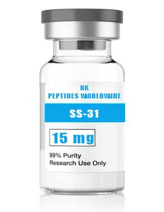
31 15 mg
Product Identity & Specifications
SS-31 (Elamipretide) 15 mg is the mid-range research format of the Szeto-Schiller series’ lead mitochondrial-targeted tetrapeptide, providing optimal material quantities for Seahorse XF metabolic flux analysis panels, moderate-throughput in vitro screening, and multi-group in vivo pilot investigations. This configuration balances per-milligram cost efficiency with practical laboratory workflow — 15 mg supports complete experimental campaigns (multiple independent biological replicates × multiple assay endpoints) from a single vial, reducing inter-batch variability that can arise when pooling material from multiple smaller vials. SS-31’s well-characterized pharmacological profile, extensive peer-reviewed literature base (>200 publications), and progression through Phase II/III clinical trials make it the most thoroughly validated mitochondrial research tool available for laboratories investigating mitochondrial quality control, cristae biology, and redox signaling.
| Specification | Detail |
|---|---|
| Peptide Name | SS-31 (Elamipretide, MTP-131, D-Arg-Dmt-Lys-Phe-NH₂) |
| Amino Acid Sequence | D-Arg-Dmt-Lys-Phe-NH₂ |
| Sequence (Single Letter) | r-Y(2,6-diMe)-k-F-NH₂ |
| CAS Registry Number | 736992-21-5 |
| Molecular Formula | C₃₂H₄₉N₉O₅ |
| Molecular Weight | 639.79 g/mol (free base); 711.91 g/mol (bis-TFA salt) |
| Number of Amino Acids | 4 (all synthetic/unusual) |
| Special Residues | D-Arg (D-arginine), Dmt (2’,6’-dimethyl-L-tyrosine) |
| C-Terminal Modification | Amide (-NH₂) |
| Purity (HPLC) | ≥98% |
| Physical Appearance | White to off-white lyophilized powder |
| Solubility | ≥5 mg/mL in H₂O, PBS, and 0.9% saline |
| Storage Condition | -20°C, desiccated, protected from light |
| Shelf Life (Lyophilized) | 24 months at recommended storage |
| Research Use Classification | Research Use Only (RUO) — Not for human or veterinary use |
Reviewed by: HKPEPTIDE WORLDWIDE Research Team | Last Updated: August 08, 2026 | Document ID: HKPW-SS31-15MG-20260808
Research Background
Mitochondrial Dysfunction as a Convergent Pathogenic Mechanism
Mitochondrial dysfunction has emerged as a convergent pathogenic node across an extraordinarily broad spectrum of human diseases — from monogenic mitochondrial disorders (Leber’s hereditary optic neuropathy, MELAS, Leigh syndrome, Barth syndrome) to highly prevalent age-associated conditions including heart failure, neurodegenerative diseases (Alzheimer’s, Parkinson’s, Huntington’s), metabolic syndrome, chronic kidney disease, and sarcopenia. The recognition that diverse etiologies converge on common mitochondrial failure mechanisms — electron transport chain dysfunction, ROS overproduction, cardiolipin peroxidation, permeability transition pore opening, and impaired mitophagy — has catalyzed intensive efforts to develop mitochondria-targeted therapeutic and research interventions (Wallace, 2005; Nunnari & Suomalainen, 2012).
Cardiolipin: The Mitochondria-Specific Lipid at the Center of Organelle Integrity
Cardiolipin occupies a privileged and unique position in eukaryotic biology as the only phospholipid synthesized exclusively within mitochondria (at the matrix face of the IMM by cardiolipin synthase, CLS) and almost entirely resident within that organelle. Its de novo biosynthesis follows an elaborate pathway: phosphatidic acid (PA) → CDP-diacylglycerol (CDP-DAG) → phosphatidylglycerol phosphate (PGP) → phosphatidylglycerol (PG) → cardiolipin (CL). Following synthesis, cardiolipin undergoes post-synthetic remodeling — the removal of saturated acyl chains and their replacement with unsaturated chains (primarily linoleic acid, C18:2) — catalyzed by tafazzin (TAZ), a transacylase whose deficiency causes Barth syndrome, an X-linked disorder characterized by dilated cardiomyopathy, skeletal myopathy, neutropenia, and 3-methylglutaconic aciduria (Schlame & Ren, 2009; Claypool & Koehler, 2012).
The Szeto-Schiller Peptides: Unintended Mitochondrial Targeting
The aromatic-cationic peptide pharmacophore that defines the SS series — alternating aromatic and basic residues in a compact structure — was originally conceived for opioid receptor targeting. The defining feature of the SS pharmacophore is the incorporation of D-amino acids and non-natural residues (Dmt — 2’,6’-dimethyltyrosine) that confer metabolic stability and optimized membrane interaction properties. The mechanism of mitochondrial targeting is elegantly simple: the combination of positive charge (from Arg/Lys) and aromatic hydrophobicity (from Phe/Dmt) enables passive diffusion across the plasma membrane, followed by electrophoretic concentration within the mitochondrial matrix driven by the substantial negative-inside membrane potential (ΔΨm ≈ -150 to -180 mV). The Nernst equation predicts approximately 1,000- to 5,000-fold accumulation for a peptide with net charge +3 (D-Arg +1, Lys +1 at pH 7.4, plus contributions from peptide termini), consistent with experimentally measured mitochondrial-to-cytosolic concentration ratios for fluorescently labeled SS-31 (Zhao et al., 2004; Szeto, 2006).
Clinical Translation and Research Validation
SS-31 has been advanced through clinical development by Stealth BioTherapeutics under the International Nonproprietary Name (INN) Elamipretide. Clinical programs have spanned mitochondrial myopathy (Phase II MMPOWER-2 trial), Barth syndrome (Phase II/III TAZPOWER), heart failure with preserved ejection fraction (HFpEF), geographic atrophy secondary to age-related macular degeneration (Phase II ReCLAIM), and primary mitochondrial disease. While clinical outcomes have been mixed — with signals of efficacy in some indications and neutral results in others — the extensive clinical dataset provides researchers with unparalleled pharmacokinetic, pharmacodynamic, and safety characterization data for a mitochondria-targeted peptide, enabling informed experimental design in preclinical research programs (Karaa et al., 2019; Couser et al., 2019; Sabbah et al., 2016).
Molecular Mechanisms
Mechanism 1: Thermodynamic and Structural Basis of Selective Cardiolipin Recognition
SS-31’s cardiolipin selectivity — >100-fold preference for cardiolipin over monobasic phospholipids — is rooted in the cooperative binding energetics enabled by cardiolipin’s unique dibasic phosphate headgroup architecture. Isothermal titration calorimetry (ITC) measurements reveal that SS-31 binding to cardiolipin-containing liposomes is exothermic (ΔH < 0) and entropy-driven at low ionic strength, consistent with electrostatic attraction as the primary energetic driver with hydrophobic burial as a contributing factor. The stoichiometry of binding is approximately 1:1 (peptide:cardiolipin), and the dissociation constant (Kd) lies in the 10⁻⁷ to 10⁻⁸ M range depending on cardiolipin acyl chain composition. Molecular dynamics simulations suggest that SS-31 adopts a hairpin-like conformation when bound to cardiolipin monolayers, with the D-Arg and Lys side chains extending toward the aqueous phase to engage phosphate headgroups while the Dmt and Phe aromatic rings penetrate approximately 5-8 Å into the acyl chain region (Birk et al., 2013; Szeto, 2014).
Mechanism 2: Interruption of the Cardiolipin-Cytochrome c Peroxidase Apoptotic Axis
The transformation of cytochrome c from an essential electron carrier to a pro-apoptotic peroxidase is one of the most dramatic functional conversions in cell biology. The molecular basis of this transformation has been elucidated through a combination of X-ray crystallography, resonance Raman spectroscopy, and stopped-flow kinetics: cardiolipin binding induces a partial unfolding of cytochrome c, particularly in the Ω-loop region (residues 70-85) housing the Met80 heme ligand. Met80 displacement from the heme iron opens the sixth coordination position for H₂O₂ binding, converting the heme center from an electron transfer protein to a peroxidatic enzyme. SS-31’s mechanism intercepts this cascade at the earliest step — by binding cardiolipin and preventing its peroxidation, it reduces the concentration of cardiolipin hydroperoxides required to induce the Met80 displacement conformational change. The net effect is preservation of cytochrome c in its native, non-peroxidatic conformation and maintenance of its physiological electron shuttle function (Kagan et al., 2005; Basova et al., 2007; Tyurina et al., 2012).
Mechanism 3: Bioenergetic Rescue — From Isolated Organelles to Intact Organisms
SS-31’s impact on mitochondrial bioenergetics has been characterized across a hierarchy of experimental systems of increasing complexity: (i) submitochondrial particles and isolated mitochondria — SS-31 preserves State 3 (ADP-stimulated) respiration and respiratory control ratio (RCR) in the presence of Complex I and Complex III inhibitors; (ii) permeabilized cells and intact cells — SS-31 maintains basal and maximal oxygen consumption rate (OCR), spare respiratory capacity, and ATP-linked respiration measured by Seahorse XF technology; (iii) isolated perfused organs — SS-31 treatment in Langendorff-perfused rat hearts subjected to ischemia-reperfusion preserves left ventricular developed pressure, reduces infarct size, and maintains myocardial ATP and phosphocreatine (PCr) levels as quantified by ³¹P-NMR spectroscopy; (iv) intact animals — SS-31 administration improves exercise tolerance, skeletal muscle mitochondrial function, and cardiac output in rodent models of heart failure, aging, and metabolic disease (Sabbah et al., 2016; Dai et al., 2011; Siegel et al., 2013).
Mechanism 4: Mitophagy Modulation and Mitochondrial Quality Control
Mitochondrial quality control operates through a coordinated network of surveillance mechanisms including mitochondrial dynamics (fusion/fission), the mitochondrial unfolded protein response (UPRmt), and selective autophagic degradation of damaged mitochondria (mitophagy). The PINK1/Parkin pathway — the best-characterized mitophagy axis — is triggered by mitochondrial depolarization, which stabilizes PINK1 on the outer mitochondrial membrane, leading to Parkin recruitment, ubiquitination of outer membrane proteins, and autophagosome engulfment. SS-31 treatment does not directly activate mitophagy but appears to reduce the fraction of mitochondria flagged for degradation by preserving ΔΨm and preventing cardiolipin externalization (an “eat-me” signal for mitophagic recognition). In aging rodent models, chronic SS-31 treatment is associated with higher mitochondrial DNA copy number, preserved mitochondrial morphology by electron microscopy, and reduced accumulation of autophagy markers (LC3-II, p62) in mitochondrial fractions — consistent with a model in which cardiolipin stabilization reduces the rate at which mitochondria become damaged beyond the threshold for mitophagic clearance (Eiyama & Okamoto, 2015; Pickles et al., 2018).
Research Applications
SS-31 15 mg enables comprehensive mitochondrial research programs:
-
Seahorse XF Bioenergetics Profiling: Complete metabolic flux analysis panels including mitochondrial stress test, glycolytic stress test, glycolytic rate assay, and ATP rate assay across multiple cell lines and treatment conditions from a single vial.
-
Moderate-Throughput Compound Screening: Screening of SS-31 across compound libraries or genetic perturbation panels (siRNA, CRISPR) for synergistic or antagonistic interactions affecting mitochondrial function.
-
Multi-Group In Vivo Pilot Studies: Three-arm pilot designs (vehicle control, SS-31 low dose, SS-31 high dose) in murine disease models with sufficient material for daily dosing and terminal tissue collection for mitochondrial functional assays.
-
Mitochondrial Proteomics and Phosphoproteomics: TMT or SILAC-based quantitative proteomics of mitochondrial-enriched fractions from SS-31-treated versus control cells/tissues, with phosphopeptide enrichment for phosphoproteomic characterization of mitochondrial signaling network changes.
-
Cristae Ultrastructure Analysis: Transmission electron microscopy (TEM) and electron tomography of mitochondrial cristae morphology in SS-31-treated tissues — the 15 mg format supports sufficient material for dose-response studies across multiple treatment groups.
-
Cardiolipin Remodeling Enzyme Studies: Investigation of SS-31 effects on cardiolipin synthase (CLS), tafazzin (TAZ), phospholipase A2 (PLA2), and monolysocardiolipin acyltransferase (MLCLAT) expression and activity.
Quality Control & Analytical Specifications
| Analytical Method | Specification | Acceptance Criteria |
|---|---|---|
| RP-HPLC Purity | C18, 214 nm UV detection | ≥98.0% peak area |
| ESI-TOF Mass Spectrometry | Positive ion mode | ⁺ 640.4 ± 0.5 Da (free base) |
| Amino Acid Analysis | Post-column ninhydrin detection | ±10% of theoretical composition |
| Peptide Content (Net Peptide) | Elemental analysis (N%) | ≥85% |
| Residual TFA | Ion chromatography | ≤1.0% |
| Water Content (Karl Fischer) | Coulometric titration | ≤8.0% w/w |
| Endotoxin | LAL kinetic chromogenic | ≤1.0 EU/mg |
| Appearance | Visual inspection | White to off-white lyophilized powder |
| Solubility | 5 mg/mL in H₂O (visual) | Clear, colorless solution |
| Chiral Purity | Chiral HPLC (Crownpak CR(+)) | ≥99% D-Arg; ≥98% L-Lys; ≥99% Dmt |
| Heavy Metal Analysis | ICP-MS | Conforms to ICH Q3D |
Available SS-31 Research Configurations
| Product Variant | Catalog Number | Quantity | Seahorse XF96 Plates (full)* | Mouse Cohorts (n=5, 5 mg/kg, 7 days) | Best Application |
|---|---|---|---|---|---|
| SS-31 5 mg | SS31-005-USA | 5 mg | 2-3 plates | — | Pilot, method validation |
| SS-31 10 mg | SS31-010-USA | 10 mg | 5-6 plates | — | Multi-endpoint mechanistic |
| SS-31 15 mg (current) | SS31-015-USA | 15 mg | 8-12 plates | ~1 cohort | Seahorse panels, multi-group in vivo |
| SS-31 30 mg | SS31-030-USA | 30 mg | 16-24 plates | ~2 cohorts | Extended in vivo, core facilities |
| Custom Bulk | SS31-BULK-USA | Inquire | — | — | Multi-institutional programs |
*Estimated at 500 nM working concentration, 100 μL per well, 1 mg/mL stock.
Frequently Asked Questions (FAQ)
1. Why choose the SS-31 15 mg format over the 10 mg format?
The SS-31 15 mg format provides 50% more material than the 10 mg configuration, making it the practical choice for laboratories conducting complete Seahorse XF metabolic flux panels. A standard mitochondrial stress test (OCR measurement with sequential oligomycin, FCCP, and rotenone/antimycin A injections) consumes approximately 1-2 mL of working solution per 96-well plate; adding a glycolytic stress test (ECAR measurement with glucose, oligomycin, and 2-DG injections) and an ATP rate assay brings the total to 3-6 mL per full panel. The 10 mg format (10 mL stock at 1 mg/mL) supports 1-2 complete panels, while the 15 mg format (15 mL stock) supports 2-4 panels with material remaining for confirmatory Western blotting or ROS assays. For in vivo work, the 15 mg format supports a complete three-group pilot study (vehicle, low dose, high dose; n=3 per group) with 5-7 days of daily dosing at 5 mg/kg in mice.
2. How many Seahorse XF assays can the 15 mg format support?
The 15 mg format reconstituted at 1 mg/mL yields 15 mL of stock solution at approximately 1.56 mM. At typical SS-31 working concentrations of 100-500 nM (final well concentration), this stock supports extensive Seahorse XF throughput. For XF96 plates (80 μL assay medium per well + 20 μL injection ports = 100 μL final volume, 500 nM SS-31): each plate requires approximately 3.2 μL of 1 mg/mL stock in 10 mL of assay medium, or approximately 0.05 mg per plate. The 15 mg vial therefore supports approximately 30 XF96 plates theoretically, though practical constraints (aliquoting, dead volume, confirmatory assays) bring the realistic number to 8-12 complete plates including biological replicates. For XF24 plates (500 μL per well), approximately 3-4 complete plates are achievable.
3. What tissue types show the greatest SS-31 accumulation following systemic administration?
SS-31 accumulates preferentially in tissues with the highest mitochondrial density and oxidative metabolic demand. Quantitative whole-body autoradiography and LC-MS/MS tissue distribution studies in rodents demonstrate the following rank order of SS-31 tissue concentrations at 30-60 minutes post-IV or IP administration: kidney cortex > heart (left ventricle) > liver > brain (cortex, hippocampus) > skeletal muscle (soleus > gastrocnemius) > lung > spleen. Tissue-to-plasma concentration ratios range from 20:1 (skeletal muscle) to 100:1 (kidney cortex), consistent with the ΔΨm-dependent electrophoretic concentration mechanism. Within the kidney, SS-31 accumulation is particularly pronounced in proximal tubular epithelial cells — the nephron segment with the highest mitochondrial density and oxidative phosphorylation dependence. In the brain, SS-31 crosses the blood-brain barrier (BBB) despite its cationic character, with brain-to-plasma ratios of approximately 5:1 to 15:1 reported depending on administration route and time point.
4. Does SS-31 affect mitochondrial biogenesis pathways?
SS-31 is not classified as a direct mitochondrial biogenesis inducer — it does not function as a PPARγ coactivator-1α (PGC-1α) agonist, PPARδ ligand, AMPK activator, or sirtuin activator (SIRT1/SIRT3). However, multiple independent laboratories have reported that SS-31 treatment under oxidative stress conditions preserves PGC-1α mRNA and protein expression levels that would otherwise decline due to oxidative damage. This preservation effect is likely indirect: by reducing mitochondrial ROS production and preventing cardiolipin peroxidation, SS-31 reduces oxidative damage to mtDNA and mitochondrial proteins, thereby maintaining a functional organelle population and reducing the cellular signal for compensatory mitochondrial biogenesis. Several studies in aging rodent models report that chronic SS-31 treatment is associated with maintained mtDNA copy number, preserved mitochondrial transcript levels (ND1, COX1, ATP6), and sustained citrate synthase activity — surrogate markers of mitochondrial content — relative to age-matched untreated controls. Researchers specifically studying mitochondrial biogenesis should consider pairing SS-31 with dedicated biogenesis inducers (AICAR, resveratrol, bezafibrate, exercise training) to investigate potential additive or synergistic effects.
5. What distinguishes SS-31 from SS-20 and other SS-family peptides?
The Szeto-Schiller peptide family comprises approximately 20 characterized aromatic-cationic peptides with systematic variations in amino acid sequence, stereochemistry, charge distribution, and aromatic residue identity. Key distinctions include: SS-02 (H-Dmt-D-Arg-Phe-Lys-NH₂) — the early lead compound from which SS-31 was derived; demonstrates mitochondrial targeting but lower cardiolipin binding affinity than SS-31. SS-20 (H-Phe-D-Arg-Phe-Lys-NH₂) — lacks the Dmt residue; exhibits mitochondrial accumulation but approximately 10-fold lower cardiolipin binding and correspondingly reduced potency in mitochondrial protection assays. SS-31 incorporates the Dmt residue at position 2, and critically, the stereochemical arrangement (D-Arg¹-Dmt²-Lys³-Phe⁴) places both the D-Arg guanidinium and Lys ε-ammonium on the same face of the peptide when in its cardiolipin-bound hairpin conformation, optimizing the bidentate electrostatic engagement geometry. This structural optimization accounts for SS-31’s superior cardiolipin binding and its status as the most extensively characterized member of the series. Researchers should note that published EC₅₀ values for mitochondrial protection often differ by an order of magnitude between SS-31 and SS-20, reflecting these structural differences.
Comparative Analysis: SS-31 Format Selection Guide
| Research Scenario | Recommended Format | Rationale |
|---|---|---|
| First-time SS-31 user, method development | 5 mg | Minimizes cost while establishing protocols |
| Routine in vitro mitochondrial assays | 10 mg | Sufficient for multi-endpoint experiments |
| Seahorse XF panels + in vitro pharmacology | 15 mg | Optimal for complete metabolic flux analysis |
| In vivo pilot studies (mouse, 5-7 days) | 15-30 mg | Supports multi-group designs |
| Chronic in vivo studies (mouse, 2-4 weeks) | 30 mg | Sufficient for sustained daily dosing |
| Core facility / multi-user environment | 30 mg or Bulk | Enables shared resource utilization |
References & Further Reading
-
Szeto, H. H. (2006). Mitochondria-targeted peptide antioxidants: Novel neuroprotective agents. AAPS Journal, 8(3), E521–E531.
-
Zhao, K., Zhao, G. M., Wu, D., Soong, Y., Birk, A. V., Schiller, P. W., & Szeto, H. H. (2004). Cell-permeable peptide antioxidants targeted to inner mitochondrial membrane inhibit mitochondrial swelling, oxidative cell death, and reperfusion injury. Journal of Biological Chemistry, 279(33), 34682–34690.
-
Birk, A. V., Liu, S., Soong, Y., Mills, W., Singh, P., Warren, J. D., Seshan, S. V., Pardee, J. D., & Szeto, H. H. (2013). The mitochondrial-targeted compound SS-31 re-energizes ischemic mitochondria by interacting with cardiolipin. Journal of the American Society of Nephrology, 24(8), 1250–1261.
-
Birk, A. V., Chao, W. M., Bracken, C., Warren, J. D., & Szeto, H. H. (2014). Targeting mitochondrial cardiolipin and the cytochrome c/cardiolipin complex to promote electron transport and optimize mitochondrial ATP synthesis. British Journal of Pharmacology, 171(8), 2017–2028.
-
Kagan, V. E., Tyurin, V. A., Jiang, J., et al. (2005). Cytochrome c acts as a cardiolipin oxygenase required for release of proapoptotic factors. Nature Chemical Biology, 1(4), 223–232.
-
Sabbah, H. N., Gupta, R. C., Kohli, S., Wang, M., Hachem, S., & Zhang, K. (2016). Chronic therapy with elamipretide (MTP-131) improves left ventricular and mitochondrial function in dogs with advanced heart failure. Circulation: Heart Failure, 9(2), e002206.
-
Claypool, S. M., & Koehler, C. M. (2012). The complexity of cardiolipin in health and disease. Trends in Biochemical Sciences, 37(1), 32–41.
-
Dai, D. F., Chen, T., Szeto, H., et al. (2011). Mitochondrial targeted antioxidant peptide ameliorates hypertensive cardiomyopathy. Journal of the American College of Cardiology, 58(1), 73–82.
-
Siegel, M. P., Kruse, S. E., Percival, J. M., et al. (2013). Mitochondrial-targeted peptide rapidly improves mitochondrial energetics and skeletal muscle performance in aged mice. Aging Cell, 12(5), 763–771.
-
Eiyama, A., & Okamoto, K. (2015). PINK1/Parkin-mediated mitophagy in mammalian cells. Current Opinion in Cell Biology, 33, 95–101.
Related Research & Resources
| Resource | Description |
|---|---|
| Glp 1 Metabolic Peptides Hub | Complete research overview & methodology hub |
| Buy Ss 31 5 Mg Research Peptide Usa | Related research peptide product |
| Ss 31 10 Mg Precision Mitochondrial Rese | Related research peptide product |
| Glp 1 Peptides Metabolic Research | Latest research insights & methodology |
View Complete Research Guide »
Compliance Statement
This SS-31 (Elamipretide) 15 mg research peptide is supplied exclusively as a Research Use Only (RUO) product for in vitro laboratory investigations and preclinical scientific research conducted within qualified research facilities. It is not manufactured in accordance with current Good Manufacturing Practice (cGMP) regulations and is not intended, approved, or labeled for human administration, veterinary therapeutic use, clinical diagnosis, treatment of any disease or medical condition, or any form of therapeutic application whatsoever. Researchers bear sole responsibility for ensuring compliance with all applicable federal, state, and institutional regulations governing laboratory chemical and peptide handling, storage, use, and disposal. Any research protocol involving this product must receive appropriate institutional oversight, including IACUC approval for in vivo research. HKPEPTIDE WORLDWIDE expressly disclaims any liability arising from improper use, misuse, diversion, or unauthorized application of this compound. By purchasing this product, the researcher acknowledges and agrees to these terms of use.
For Certificate of Analysis requests, technical inquiries, or institutional procurement, contact the HKPEPTIDE WORLDWIDE Research Support Team.