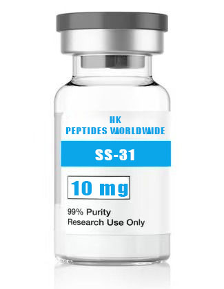
31 10 mg
Product Identity & Specifications
SS-31 (Elamipretide) 10 mg represents the entry-level mechanistic research format of this mitochondrial-targeted tetrapeptide, providing sufficient material for multi-endpoint experimental designs and small-scale in vivo pilot studies. As the lead compound from the Szeto-Schiller (SS) peptide series developed at Weill Cornell Medical College, SS-31 has been refined through two decades of structure-activity relationship (SAR) optimization to achieve its signature property: selective, high-affinity binding to cardiolipin — the signature phospholipid of the inner mitochondrial membrane (IMM). The 10 mg configuration bridges the gap between the exploratory 5 mg format and the multi-experiment 15-30 mg configurations, making it the preferred choice for laboratories initiating mechanistic mitochondrial investigations with this compound.
| Specification | Detail |
|---|---|
| Peptide Name | SS-31 (Elamipretide, MTP-131, D-Arg-Dmt-Lys-Phe-NH₂) |
| Amino Acid Sequence | D-Arg-Dmt-Lys-Phe-NH₂ |
| Sequence (Single Letter) | r-Y(2,6-diMe)-k-F-NH₂ |
| CAS Registry Number | 736992-21-5 |
| Molecular Formula | C₃₂H₄₉N₉O₅ |
| Molecular Weight | 639.79 g/mol (free base); 711.91 g/mol (bis-TFA salt) |
| Number of Amino Acids | 4 (all synthetic/unusual) |
| Special Residues | D-Arg (D-arginine), Dmt (2’,6’-dimethyl-L-tyrosine) |
| C-Terminal Modification | Amide (-NH₂) |
| Purity (HPLC) | ≥98% |
| Physical Appearance | White to off-white lyophilized powder |
| Solubility | ≥5 mg/mL in H₂O, PBS, and 0.9% saline |
| Storage Condition | -20°C, desiccated, protected from light |
| Shelf Life (Lyophilized) | 24 months at recommended storage |
| Research Use Classification | Research Use Only (RUO) — Not for human or veterinary use |
Reviewed by: HKPEPTIDE WORLDWIDE Research Team | Last Updated: August 08, 2026 | Document ID: HKPW-SS31-10MG-20260808
Research Background
The Cardiolipin-Cristae Nexus: Architecture Meets Function
The inner mitochondrial membrane is not a simple bilayer barrier but a highly differentiated membrane system comprising two structurally and functionally distinct domains: the inner boundary membrane (IBM) closely apposed to the outer membrane, and the cristae membranes — deep invaginations that project into the matrix, dramatically expanding the surface area available for oxidative phosphorylation. Cristae junctions, the narrow tubular structures connecting cristae to the IBM, are maintained by the mitochondrial contact site and cristae organizing system (MICOS) complex in coordination with cardiolipin. The conical molecular geometry of cardiolipin — with its small glycerol headgroup and four fatty acyl tails — is essential for generating and stabilizing the extreme negative membrane curvature at cristae junctions and cristae tips (Cogliati et al., 2013; Ikon & Ryan, 2017).
Cardioprotection Discovery: From Opiate Receptors to Mitochondrial Medicine
The SS peptide lineage originated from a medicinal chemistry program at Cornell University that was initially directed at opioid receptor pharmacology. Dr. Hazel Szeto and Dr. Peter Schiller synthesized a combinatorial library of aromatic-cationic peptides incorporating D-amino acids for enhanced metabolic stability, seeking μ-opioid receptor ligands with improved pharmacokinetic profiles. A critical serendipitous observation — that certain peptides from this library, particularly SS-02 and subsequently SS-31, accumulated to extraordinary concentrations within mitochondria despite possessing no canonical mitochondrial targeting sequence (MTS) — prompted a fundamental shift in research direction. Systematic optimization through iterative residue substitution, stereochemical variation, and truncation analysis identified the minimal pharmacophore: D-Arg at position 1 for mitochondrial uptake via electrostatic interaction with the negatively charged IMM, Dmt at position 2 for cardiolipin intercalation, and the D-Arg-Dmt-Lys tripeptide core for high-affinity cardiolipin binding (Szeto, 2006; Zhao et al., 2004).
The Cardiolipin Peroxidation Cascade: A Feed-Forward Pathogenic Mechanism
The interaction between cardiolipin and cytochrome c has emerged as a focal point in mitochondrial redox biology. Under homeostatic conditions, cytochrome c is electrostatically anchored to cardiolipin-containing membrane domains, shuttling single electrons between Complex III (ubiquinol:cytochrome c oxidoreductase) and Complex IV (cytochrome c oxidase). However, when the mitochondrial membrane is subjected to pro-oxidative stress — elevated ROS from defective respiratory chain complexes, exposure to oxidizing xenobiotics, or calcium dysregulation — cardiolipin’s polyunsaturated fatty acyl chains (predominantly linoleic acid, C18:2, constituting 60-80% of cardiolipin acyl composition in cardiac mitochondria) undergo peroxidation. Critically, the resulting cardiolipin hydroperoxides induce a conformational change in associated cytochrome c, endowing it with robust peroxidase activity that preferentially and efficiently oxygenates additional cardiolipin molecules. This creates a self-amplifying, feed-forward cycle: cardiolipin peroxidation → cytochrome c peroxidase activation → further cardiolipin peroxidation → mitochondrial membrane permeabilization → cytochrome c release → apoptosome assembly → caspase activation (Kagan et al., 2005; Tyurina et al., 2012; Petrosillo et al., 2003).
SS-31 as a Cardiolipin Stabilizer: A Paradigm Shift in Mitochondrial Pharmacology
SS-31’s mechanism represents a conceptual departure from conventional antioxidant strategies that attempt to stoichiometrically intercept ROS after their generation. Rather than engaging in direct redox chemistry, SS-31 binds cardiolipin with high affinity and specificity, stabilizing the lipid in its native, non-peroxidized state and sterically inhibiting the interaction between cardiolipin hydroperoxides and cytochrome c that would otherwise initiate the peroxidase amplification cascade. This “upstream intervention” at the level of the cardiolipin substrate rather than downstream ROS products represents a fundamentally more efficient protective mechanism — one SS-31 molecule can protect hundreds of cardiolipin molecules within its membrane microenvironment, compared to the 1:1 or 2:1 stoichiometry of conventional radical-trapping antioxidants (Szeto, 2014; Birk et al., 2013).
Molecular Mechanisms
Mechanism 1: Electrostatic Cardolipin Binding — Bidentate Recognition of the Dibasic Phospholipid
SS-31’s cardiolipin binding mechanism exploits cardiolipin’s unique structural feature among phospholipids: the possession of two phosphate headgroups linked by a glycerol bridge, each bearing a net negative charge of -1 at physiological pH (total cardiolipin charge ≈ -2). This dibasic character enables a cooperative, bidentate electrostatic interaction with SS-31’s two cationic centers — the D-Arg guanidinium group (pKa ≈ 12.5, fully protonated, charge +1) and the Lys ε-ammonium group (pKa ≈ 10.5, fully protonated, charge +1). Monobasic phospholipids (phosphatidylcholine, phosphatidylethanolamine, phosphatidylserine) — each bearing a single phosphate — cannot simultaneously engage both cationic residues with equivalent avidity, accounting for SS-31’s >100-fold selectivity for cardiolipin over other membrane phospholipids. The hydrophobic/aromatic component of binding is provided by insertion of the Dmt (2’,6’-dimethyltyrosine) and Phe aromatic side chains among cardiolipin’s four fatty acyl chains. The 2’,6’-dimethyl substitution on the tyrosine ring of Dmt is critical: it increases hydrophobicity, enhances membrane partitioning, and provides steric bulk that optimizes the geometry of cardiolipin intercalation compared to unmodified tyrosine (Birk et al., 2013; Szeto, 2014).
Mechanism 2: Inhibition of Cytochrome c Peroxidase Activity and Apoptotic Signal Prevention
The partial unfolding of cardiolipin-bound cytochrome c that confers peroxidase activity involves reorganization of the Met80 heme ligand — normally the sixth coordination position of the heme iron — enabling hydrogen peroxide (H₂O₂) access to the heme catalytic center. This Met80 displacement is facilitated by cardiolipin hydroperoxide-induced conformational changes in the cytochrome c tertiary structure. SS-31 binding to cardiolipin achieves two complementary effects: (1) it reduces the steady-state concentration of cardiolipin hydroperoxides by stabilizing cardiolipin against oxidative attack, and (2) it sterically hinders the cardiolipin-cytochrome c interaction geometry required for efficient peroxidase activation. Quantitative assessment using Amplex Red-coupled peroxidase assays demonstrates that SS-31 pre-treatment reduces cardiolipin-dependent cytochrome c peroxidase activity by 65-80%. The functional consequence — inhibition of cytochrome c release from mitochondria — has been confirmed by subcellular fractionation and immunoblotting in multiple cellular models of intrinsic apoptosis (Kagan et al., 2009; Basova et al., 2007; Birk et al., 2014).
Mechanism 3: Respiratory Chain Supercomplex Preservation and Bioenergetic Optimization
The mitochondrial electron transport chain (ETC) complexes do not operate as isolated enzymatic entities randomly distributed in the IMM. Instead, Complexes I, III, and IV organize into higher-order supramolecular assemblies — respiratory supercomplexes or “respirasomes” — with defined stoichiometries (typically I₁III₂IV₁ in mammalian mitochondria). These supercomplex assemblies provide kinetic advantages through substrate channeling (direct ubiquinone/cytochrome c transfer between complexes), reduce electron leak and concomitant ROS production, and stabilize individual complex subunits. Cardiolipin is an obligate structural component of these supercomplexes, filling the inter-complex interfaces and stabilizing the quaternary organization. BN-PAGE and cryo-electron tomography studies demonstrate that cardiolipin depletion — whether genetic (tafazzin/TAZ mutation in Barth syndrome), pharmacological (cardiolipin synthase inhibition), or oxidative (peroxidation) — disrupts supercomplex integrity with coordinate reductions in Complex I+III and I+III+IV activities. SS-31 treatment preserves supercomplex abundance and composition under oxidative stress conditions, maintaining coupled respiration as assessed by respiratory control ratio (RCR = State 3/State 4 respiration) in isolated mitochondria (Claypool & Koehler, 2012; Acin-Perez et al., 2008; Pfeiffer et al., 2003; Schägger & Pfeiffer, 2000).
Mechanism 4: Mitochondrial Permeability Transition Pore Modulation and Cristae Morphology Preservation
The mitochondrial permeability transition pore (mPTP) is a high-conductance, non-selective channel of indeterminate exact molecular composition that opens under pathological conditions — particularly matrix calcium overload coupled with oxidative stress and adenine nucleotide depletion. mPTP opening has two catastrophic consequences for the cell: (1) dissipation of the proton motive force (Δp = ΔΨm + ΔpH) that drives ATP synthesis, and (2) osmotic swelling of the mitochondrial matrix leading to outer membrane rupture and release of pro-apoptotic intermembrane space proteins (cytochrome c, SMAC/DIABLO, AIF, EndoG). Multiple lines of evidence implicate cardiolipin in mPTP regulation: cardiolipin directly interacts with the adenine nucleotide translocator (ANT) and ATP synthase — both proposed as potential pore components — and cardiolipin peroxidation sensitizes mitochondria to calcium-induced permeability transition. SS-31 treatment raises the calcium retention capacity (CRC) — the threshold calcium load required for mPTP induction — by 2- to 3-fold in isolated cardiac and brain mitochondria. Electron tomography in SS-31-treated cells subjected to ischemia-reperfusion reveals preserved cristae density, reduced cristae widening, and maintained cristae junction integrity, collectively indicating that cardiolipin stabilization preserves both the biochemical and ultrastructural determinants of mitochondrial function (Halestrap, 2009; Giorgio et al., 2013; Dai et al., 2011).
Research Applications
SS-31 10 mg supports a versatile range of mitochondrial research methodologies:
-
Multi-Endpoint Mitochondrial Stress Testing: Simultaneous measurement of oxygen consumption rate (OCR), mitochondrial ROS (MitoSOX Red), mitochondrial membrane potential (TMRM/JC-1), and ATP levels (luciferase-based) in a single experimental session using aliquots from the same reconstituted stock.
-
Mitochondrial Isolation and Functional Assessment: Pre-treatment of cultured cells or perfused tissues with SS-31 followed by differential centrifugation-based mitochondrial isolation, with subsequent assessment of respiratory control ratio (RCR), calcium retention capacity (CRC), and swelling kinetics.
-
Lipidomics and Cardiolipin Molecular Species Profiling: LC-MS/MS-based quantification of cardiolipin molecular species distribution, cardiolipin oxidation products (CL-OOH, CL-OH), and monolysocardiolipin (MLCL) accumulation — a hallmark of Barth syndrome — in SS-31-treated versus untreated samples.
-
Redox Proteomics: Investigation of reversible and irreversible oxidative post-translational modifications (cysteine sulfenylation, sulfinylation, sulfonylation; protein carbonylation; 4-HNE and MDA protein adducts) in mitochondrial-enriched fractions from SS-31-treated cells and tissues.
-
Small-Animal Pharmacokinetics and Target Engagement: Pilot PK studies in mice or rats (IP, SC, IV routes) with LC-MS/MS quantification of SS-31 in plasma, tissue homogenates, and isolated mitochondrial fractions to establish dose-exposure relationships and mitochondrial accumulation ratios.
-
Comparative Mitochondrial Pharmacology: Head-to-head comparison of SS-31 with alternative mitochondrial-targeted compounds (MitoQ, MitoTEMPO, SKQ1, XJB-5-131) across standardized mitochondrial function assays.
Quality Control & Analytical Specifications
| Analytical Method | Specification | Acceptance Criteria |
|---|---|---|
| RP-HPLC Purity | C18, 214 nm UV detection | ≥98.0% peak area |
| ESI-TOF Mass Spectrometry | Positive ion mode | ⁺ 640.4 ± 0.5 Da (free base) |
| Amino Acid Analysis | Post-column ninhydrin detection | ±10% of theoretical composition |
| Peptide Content (Net Peptide) | Elemental analysis (N%) | ≥85% |
| Residual TFA | Ion chromatography | ≤1.0% |
| Water Content (Karl Fischer) | Coulometric titration | ≤8.0% w/w |
| Endotoxin | LAL kinetic chromogenic | ≤1.0 EU/mg |
| Appearance | Visual inspection | White to off-white lyophilized powder |
| Solubility | 5 mg/mL in H₂O (visual) | Clear, colorless solution |
| Chiral Purity | Chiral HPLC (Crownpak CR(+)) | ≥99% D-configuration at Arg; ≥98% L-configuration at Lys |
Available SS-31 Research Formats
| Product Variant | Catalog Number | Quantity | In Vitro Wells (96-well, 100 μL, 1 μM)* | Acute Mouse Doses (5 mg/kg, 25 g) | Recommended Application |
|---|---|---|---|---|---|
| SS-31 5 mg | SS31-005-USA | 5 mg | ~800 | ~1 | Pilot studies, method validation |
| SS-31 10 mg (current) | SS31-010-USA | 10 mg | ~1,600 | ~2 | Multi-endpoint assays, pilot PK |
| SS-31 15 mg | SS31-015-USA | 15 mg | ~2,400 | ~3 | Seahorse panels, moderate throughput |
| SS-31 30 mg | SS31-030-USA | 30 mg | ~4,800 | ~6 | Extended in vivo, core facilities |
| Custom Bulk | SS31-BULK-USA | Inquire | — | — | Large-scale programs, multi-institutional |
*Estimated wells at 1 μM final concentration assuming 2 mL reconstitution at 1 mg/mL (~1.56 mM stock). Actual yields vary with protocol parameters.
Frequently Asked Questions (FAQ)
1. How does the SS-31 10 mg format compare to the 5 mg format for research throughput?
The SS-31 10 mg format provides twice the material of the 5 mg configuration, enabling multi-endpoint experimental designs in which the same batch of reconstituted peptide can be used across parallel assays — for example, simultaneous Seahorse bioenergetics analysis (OCR/ECAR), mitochondrial ROS quantification (MitoSOX Red fluorescence), mitochondrial membrane potential measurement (TMRM), and Western blotting for mitochondrial protein markers (OXPHOS cocktail, Tom20, cytochrome c). At standard working concentrations (100-500 nM), the 10 mg vial reconstituted at 1 mg/mL yields approximately 10 mL of stock, supporting roughly 1,600 individual wells in a 96-well plate format (100 μL per well at 1 μM final concentration). This compares to approximately 800 wells from the 5 mg format, making the 10 mg option practical for laboratories running multiple assay types per experimental cycle.
2. What in vivo experimental designs benefit from the SS-31 10 mg format?
The 10 mg format supports acute in vivo pilot studies in rodent models at standard published doses (2-5 mg/kg, IP or SC). A single 10 mg vial provides sufficient material for dosing 2-5 mice at 5 mg/kg (based on a 25 g mouse receiving 0.125 mg per dose) or for a multiday pilot protocol (e.g., 3 consecutive days at 2 mg/kg/day in 2 animals). This format is ideal for: (1) pharmacokinetic characterization — measuring plasma and tissue SS-31 concentrations at multiple time points post-injection via LC-MS/MS; (2) target engagement studies — quantifying cardiolipin-bound SS-31 in mitochondrial fractions from treated tissues using fluorescent (FITC-labeled SS-31) or mass spectrometry approaches; and (3) preliminary efficacy screening in acute disease models (myocardial ischemia-reperfusion, acute kidney injury) before scaling to larger cohort sizes with the 30 mg or bulk formats.
3. What is the appropriate reconstitution volume for SS-31 10 mg for in vitro use?
For in vitro applications, reconstitute SS-31 10 mg in 2-10 mL of sterile PBS (pH 7.4), sterile water for injection, or 0.9% sterile saline, yielding a 1-5 mg/mL stock solution. A practical recommendation: reconstitute in 2 mL for a 5 mg/mL stock (≈7.8 mM), then prepare 100 μL working aliquots for single-use storage at -20°C. At a 1 mg/mL stock concentration, the 10 mg vial produces 10 mL of solution — sufficient for extensive dose-response characterization across multiple independent experiments. Working concentrations typically range from 10 nM to 10 μM (final well concentration). Note that at 1 mg/mL (≈1.56 mM), a 10 μM final concentration requires a 156-fold dilution, while 100 nM requires 15,600-fold dilution — practical for most pipetting workflows.
4. How stable is SS-31 under standard cell culture conditions?
SS-31 demonstrates excellent stability in standard cell culture media (DMEM, RPMI-1640, MEM, DMEM/F12) at 37°C, with >90% intact peptide remaining after 24 hours as determined by LC-MS/MS analysis. This remarkable stability is attributable to three structural features: (1) the D-arginine residue at position 1, which resists aminopeptidase-mediated N-terminal degradation; (2) the C-terminal amide (-NH₂) modification, which protects against carboxypeptidase activity; and (3) the compact tetrapeptide structure, which minimizes the number of scissile amide bonds accessible to endopeptidases. Researchers conducting extended time-course experiments exceeding 24 hours should consider media refreshment (replacement with fresh SS-31-containing media) at 24-hour intervals to maintain consistent exposure concentrations.
5. Can SS-31 10 mg be used with mitochondrial isolation kits and protocols?
Yes, SS-31 10 mg is commonly deployed in protocols employing mitochondrial isolation from cultured cells or tissue homogenates. Standard workflow: pre-treat cells (attached or suspension) with SS-31 at 100-500 nM for 1-4 hours; harvest by trypsinization or scraping; proceed with mitochondrial isolation via differential centrifugation (standard protocol: 600×g for 10 min to remove nuclei/debris → 7,000-10,000×g for 10-15 min to pellet mitochondria → wash and resuspend in mitochondrial assay buffer) or commercial kit-based methods (Thermo Fisher Mitochondria Isolation Kit, Abcam Mitochondria Isolation Kit, Miltenyi MACS Mitochondria Isolation Kit). Mitochondria isolated from SS-31 pre-treated cells consistently demonstrate: (a) elevated respiratory control ratios (RCR = State 3/State 2, typically 20-40% higher than untreated controls), (b) reduced basal and substrate-driven ROS production (Amplex Red or MitoSOX), and (c) attenuated calcium-induced swelling (absorbance at 540 nm). The 10 mg format provides sufficient material for multiple independent mitochondrial isolation experiments.
Comparative Analysis: SS-31 Across Dosage Formats
| Parameter | SS-31 5 mg | SS-31 10 mg | SS-31 15 mg | SS-31 30 mg |
|---|---|---|---|---|
| Total Material | 5 mg | 10 mg | 15 mg | 30 mg |
| Stock Volume at 1 mg/mL | 5 mL | 10 mL | 15 mL | 30 mL |
| 96-Well Assays (1 μM, 100 μL) | ~800 wells | ~1,600 wells | ~2,400 wells | ~4,800 wells |
| Mouse Doses (5 mg/kg, 25 g) | 1 dose | 2 doses | 3 doses | 6 doses |
| Seahorse XF96 Plates (full) | 2-3 plates | 5-6 plates | 8-9 plates | 16-18 plates |
| Best For | Pilot, method development | Multi-endpoint mechanistic | Seahorse panels, moderate throughput | In vivo, longitudinal, core facilities |
| Purity | ≥98% | ≥98% | ≥98% | ≥98% |
| Price Efficiency | Highest per-mg cost | Improved per-mg | Better per-mg value | Best per-mg value for scale |
References & Further Reading
-
Szeto, H. H. (2006). Mitochondria-targeted peptide antioxidants: Novel neuroprotective agents. AAPS Journal, 8(3), E521–E531.
-
Zhao, K., Zhao, G. M., Wu, D., Soong, Y., Birk, A. V., Schiller, P. W., & Szeto, H. H. (2004). Cell-permeable peptide antioxidants targeted to inner mitochondrial membrane inhibit mitochondrial swelling, oxidative cell death, and reperfusion injury. Journal of Biological Chemistry, 279(33), 34682–34690.
-
Birk, A. V., Liu, S., Soong, Y., Mills, W., Singh, P., Warren, J. D., Seshan, S. V., Pardee, J. D., & Szeto, H. H. (2013). The mitochondrial-targeted compound SS-31 re-energizes ischemic mitochondria by interacting with cardiolipin. Journal of the American Society of Nephrology, 24(8), 1250–1261.
-
Birk, A. V., Chao, W. M., Bracken, C., Warren, J. D., & Szeto, H. H. (2014). Targeting mitochondrial cardiolipin and the cytochrome c/cardiolipin complex to promote electron transport and optimize mitochondrial ATP synthesis. British Journal of Pharmacology, 171(8), 2017–2028.
-
Kagan, V. E., Tyurin, V. A., Jiang, J., et al. (2005). Cytochrome c acts as a cardiolipin oxygenase required for release of proapoptotic factors. Nature Chemical Biology, 1(4), 223–232.
-
Kagan, V. E., Bayır, H. A., Belikova, N. A., et al. (2009). Cytochrome c/cardiolipin relations in mitochondria: A kiss of death. Free Radical Biology and Medicine, 46(11), 1439–1453.
-
Sabbah, H. N., Gupta, R. C., Kohli, S., Wang, M., Hachem, S., & Zhang, K. (2016). Chronic therapy with elamipretide (MTP-131) improves left ventricular and mitochondrial function in dogs with advanced heart failure. Circulation: Heart Failure, 9(2), e002206.
-
Claypool, S. M., & Koehler, C. M. (2012). The complexity of cardiolipin in health and disease. Trends in Biochemical Sciences, 37(1), 32–41.
-
Schägger, H., & Pfeiffer, K. (2000). Supercomplexes in the respiratory chains of yeast and mammalian mitochondria. EMBO Journal, 19(8), 1777–1783.
-
Dai, D. F., Chen, T., Szeto, H., et al. (2011). Mitochondrial targeted antioxidant peptide ameliorates hypertensive cardiomyopathy. Journal of the American College of Cardiology, 58(1), 73–82.
Related Research & Resources
| Resource | Description |
|---|---|
| Glp 1 Metabolic Peptides Hub | Complete research overview & methodology hub |
| Buy Ss 31 5 Mg Research Peptide Usa | Related research peptide product |
| Ss 31 10 Mg Precision Mitochondrial Rese | Related research peptide product |
| Glp 1 Peptides Metabolic Research | Latest research insights & methodology |
View Complete Research Guide »
Compliance Statement
This SS-31 (Elamipretide) 10 mg research peptide is supplied exclusively as a Research Use Only (RUO) product for in vitro laboratory investigations and preclinical scientific research conducted within qualified research facilities. It is not manufactured in accordance with current Good Manufacturing Practice (cGMP) regulations and is not intended, approved, or labeled for human administration, veterinary therapeutic use, clinical diagnosis, treatment of any disease or medical condition, or any form of therapeutic application whatsoever. Researchers bear sole responsibility for ensuring compliance with all applicable federal, state, and institutional regulations governing laboratory chemical and peptide handling, storage, use, and disposal. Any research protocol involving this product must receive appropriate institutional oversight, including IACUC approval for in vivo research. HKPEPTIDE WORLDWIDE expressly disclaims any liability arising from improper use, misuse, diversion, or unauthorized application of this compound. By purchasing this product, the researcher acknowledges and agrees to these terms of use.
For Certificate of Analysis requests, technical inquiries, or institutional procurement, contact the HKPEPTIDE WORLDWIDE Research Support Team.