NAD+ (Nicotinamide Adenine Dinucleotide, Oxi
Coenzyme & Cellular Metabolism ResearchReviewed by: HKPEPTIDE WORLDWIDE Research Team | Last Updated: 2026-08-08 | Document ID: HKPW-nad-plus-research-grade-nicotinamide-adenine-dinucleotide-v1.0
1. Product Identity & Specifications
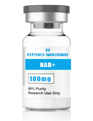
Nicotinamide Adenine Dinucleotide (NAD+, oxidized form) is the central coenzyme of cellular metabolism, functioning as both an obligate electron carrier in oxidative phosphorylation and a consumable substrate for three major enzyme families: sirtuins (class III histone deacetylases), poly(ADP-ribose) polymerases (PARPs), and cyclic ADP-ribose synthases (CD38, CD157). HKPEPTIDE WORLDWIDE supplies high-purity NAD+ (≥98% by HPLC) in three configurations—100 mg, 500 mg, and 1000 mg—spanning the range from pilot assay development to institutional-scale research programs.
NAD+ is composed of two nucleotides (adenosine monophosphate and nicotinamide ribotide) joined through their 5’-phosphate groups by a pyrophosphate (P-O-P) linkage. The nicotinamide moiety undergoes reversible hydride (H⁻) transfer at the para position of the pyridinium ring—the biochemical basis for NAD+‘s role as an electron acceptor. The oxidized form (NAD+) absorbs at 260 nm (ε = 18,000 M⁻¹cm⁻¹) and is non-fluorescent; the reduced form (NADH) additionally absorbs at 340 nm (ε = 6,220 M⁻¹cm⁻¹) and emits fluorescence at 460 nm—properties widely exploited for continuous enzyme-coupled assays.
| Parameter | Specification |
|---|---|
| Product Name | β-Nicotinamide Adenine Dinucleotide (NAD+, Oxidized Form) |
| CAS Number | 53-84-9 |
| Molecular Formula | C₂₁H₂₇N₇O₁₄P₂ |
| Molecular Weight | 663.4 Da (free acid) |
| Available Configurations | 100 mg, 500 mg, 1000 mg |
| Appearance | White to off-white lyophilized powder |
| Purity | ≥98% by RP-HPLC (C18, 260 nm detection) |
| UV Absorption | λmax = 260 nm; A₂₅₀/A₂₆₀ = 0.83 ± 0.02; A₂₈₀/A₂₆₀ = 0.22 ± 0.02 |
| Solubility | ≥100 mg/mL in water; soluble in PBS (pH 7.0–7.4) |
| Storage (Lyophilized) | -20°C, desiccated, protected from light and moisture |
| Storage (Reconstituted) | Aliquot; store at -80°C; stable ≥6 months under these conditions |
| Product Grade | Research Use Only (RUO) |
| Hygroscopicity | Highly hygroscopic — handle under dry atmosphere; minimize ambient exposure |
2. Research Background
NAD+ was first identified by Harden and Young in 1906 as a heat-stable, dialyzable cofactor required for alcoholic fermentation in yeast extracts—a discovery that earned Harden the 1929 Nobel Prize in Chemistry. The complete chemical structure was elucidated by von Euler-Chelpin (Nobel Prize, 1929) and Warburg in the 1930s, establishing NAD+ as the first coenzyme to be structurally characterized. The identification of NAD+‘s role in biological oxidations by Warburg and its function in glycolysis by Embden, Meyerhof, and Parnas cemented NAD+ as a cornerstone molecule of biochemistry.
For most of the 20th century, NAD+ was understood primarily through its canonical redox function: shuttling electrons between metabolic pathways via the NAD+/NADH couple. This redox-centric view was fundamentally expanded by the discoveries of Sir2 (the founding sirtuin) as a NAD+-dependent histone deacetylase (Guarente laboratory, 2000), PARP-1 as a NAD+-consuming DNA damage sensor (Chambon laboratory, 1963; de Murcia, 1990s), and CD38 as a multifunctional NAD+ glycohydrolase and ADP-ribosyl cyclase (Howard laboratory, 1993). These discoveries revealed NAD+ as a consumable signaling molecule whose intracellular concentration governs fundamental processes including gene silencing, DNA repair, calcium signaling, and circadian rhythm.
The biological significance of NAD+ extends across virtually every area of biomedical research. In aging biology, NAD+ levels decline by up to 50% between ages 40 and 70 in humans, and this decline correlates with age-associated mitochondrial dysfunction, genomic instability, and loss of stem cell regenerative capacity—a relationship that has driven intense interest in NAD+ augmentation strategies using precursors (NR, NMN) and NAD+-consuming enzyme inhibitors (CD38 inhibitors, PARP inhibitors). In metabolic disease research, NAD+ regulates hepatic gluconeogenesis, adipocyte lipolysis, and pancreatic β-cell insulin secretion through SIRT1-dependent deacetylation of PGC-1α, FOXO1, and UCP2. In neurodegeneration research, NAD+ depletion is a common pathological feature in models of Alzheimer’s disease, Parkinson’s disease, and axonal degeneration—the latter mediated by the NAD+-consuming Wallerian degeneration enzyme SARM1.
For the research community, HKPEPTIDE WORLDWIDE’s three-tier NAD+ configuration addresses the full spectrum of experimental scales: 100 mg for individual investigator-led assay development; 500 mg for multi-arm pharmacological studies and core facility stocks; 1000 mg for institutional research programs and large-scale biochemical assays.
3. Molecular Mechanisms
3.1 Redox Chemistry: The NAD+/NADH Couple
The defining biochemical property of NAD+ is its ability to reversibly accept a hydride ion (H⁻ = H⁺ + 2e⁻) at the C4 position of the nicotinamide ring, converting to NADH. This transfer is stereospecific: dehydrogenases are classified as A-type (pro-R hydrogen transferred, e.g., alcohol dehydrogenase) or B-type (pro-S hydrogen transferred, e.g., glyceraldehyde-3-phosphate dehydrogenase). The standard reduction potential of the NAD+/NADH couple is -320 mV (at pH 7.0, 25°C), positioning NADH as a strong reductant capable of donating electrons to Complex I (NADH:ubiquinone oxidoreductase) of the mitochondrial electron transport chain—the entry point for the majority of cellular oxygen consumption.
In the cytosol, the NAD+/NADH ratio is typically 500–700:1, favoring NAD+-dependent oxidative reactions (glycolysis, the malate-aspartate shuttle). In the mitochondrial matrix, the ratio is 5–10:1, reflecting the NADH-rich environment necessary for oxidative phosphorylation. These compartment-specific ratios are maintained by the inner mitochondrial membrane’s impermeability to NAD(H) and the operation of shuttle systems (malate-aspartate and glycerol-3-phosphate) that transfer reducing equivalents without net NAD(H) transport.
3.2 Sirtuin Activation: NAD+-Dependent Deacetylation
Sirtuins (SIRT1–7) are class III histone deacetylases that couple lysine deacetylation to NAD+ hydrolysis, producing nicotinamide (NAM), 2’-O-acetyl-ADP-ribose (OAADPr), and the deacetylated substrate. The catalytic mechanism proceeds through a unique ADP-ribosyltransferase-like intermediate: the nicotinamide moiety of NAD+ is cleaved, generating an oxocarbenium ion at C1’ of the ribose ring, which is captured by the acetyl oxygen of the acetyl-lysine substrate to form a 1’-O-alkylamidate intermediate. This intermediate undergoes base-catalyzed decomposition to yield OAADPr and the deacetylated lysine.
Sirtuin activity is directly governed by NAD+ availability. SIRT1, the most extensively studied isoform, has a Km for NAD+ of approximately 150–200 μM—values that lie within the range of physiological NAD+ fluctuations, making SIRT1 an exquisite sensor of cellular energy status. Key SIRT1 substrates include: PGC-1α (deacetylation activates mitochondrial biogenesis), FOXO1/FOXO3a (deacetylation promotes antioxidant gene expression and stress resistance), p53 (deacetylation suppresses pro-apoptotic function), NF-κB p65 (deacetylation reduces inflammatory gene transcription), and histone H3 (H3K9ac, H3K14ac deacetylation represses repetitive elements and promotes genomic stability).
3.3 PARP Activation: NAD+ as a DNA Damage Sensor Substrate
PARP-1, PARP-2, and tankyrases (PARP-5a/5b) catalyze the transfer of ADP-ribose units from NAD+ to acceptor proteins, building linear or branched poly(ADP-ribose) (PAR) chains. PARP-1, the founding family member, is activated by binding to DNA single-strand and double-strand breaks, whereupon it synthesizes massive PAR chains on itself (auto-PARylation), histones, and DNA repair proteins. This PARylation serves as a scaffold recruiting XRCC1, DNA ligase III, and other base excision repair (BER) factors to damage sites.
PARP-1 activation is the single largest consumer of cellular NAD+ under conditions of genotoxic stress. Severe DNA damage (e.g., from alkylating agents, ionizing radiation, or oxidative stress) causes hyperactivation of PARP-1, depleting cellular NAD+ to less than 20% of basal levels within minutes and triggering ATP depletion, energetic collapse, and a distinct form of programmed necrosis termed parthanatos. This NAD+ depletion/PARthanatos axis is implicated in ischemia-reperfusion injury, excitotoxicity, and inflammatory tissue damage—making PARP inhibitors and NAD+ repletion strategies complementary therapeutic research directions.
3.4 CD38 and NAD+ Glycohydrolase Activity
CD38 (and its homolog CD157/BST-1) are multifunctional ectoenzymes that consume NAD+ to generate three second messengers: cyclic ADP-ribose (cADPR, a Ca²⁺-mobilizing agent), ADP-ribose (ADPR, a TRPM2 channel agonist), and nicotinic acid adenine dinucleotide phosphate (NAADP, the most potent Ca²⁺-mobilizing second messenger known). CD38 is the predominant NAD+-consuming enzyme in mammalian tissues and is largely responsible for the age-associated decline in tissue NAD+ levels. CD38 expression increases with age and is induced by inflammatory cytokines and cellular senescence via the transcription factor IRF1. CD38 knockout mice maintain youthful NAD+ levels into old age, and pharmacological CD38 inhibition with apigenin, quercetin, or the tool compound 78c elevates tissue NAD+ by 10–30-fold—findings that have catalyzed intense interest in CD38 as a therapeutic target for age-associated metabolic decline.
3.5 NAD+ Biosynthesis: Salvage, Preiss-Handler, and De Novo Pathways
Mammalian cells maintain NAD+ pools through three biosynthetic routes. The salvage pathway (predominant in most tissues): nicotinamide (NAM) → nicotinamide mononucleotide (NMN) catalyzed by nicotinamide phosphoribosyltransferase (NAMPT, the rate-limiting enzyme) → NAD+ via NMN adenylyltransferases (NMNAT1–3). The Preiss-Handler pathway: nicotinic acid (NA) → nicotinic acid mononucleotide (NAMN) via NAPRT → nicotinic acid adenine dinucleotide (NAAD) via NMNATs → NAD+ via NAD+ synthetase (NADSYN1). The de novo pathway: tryptophan → quinolinic acid (via the kynurenine pathway) → NAMN → NAD+.
NAMPT is the principal regulator of mammalian NAD+ biosynthesis under basal conditions. NAMPT expression and activity are regulated by the circadian clock (CLOCK:BMAL1 → NAMPT → SIRT1 → BMAL1 deacetylation, a transcriptional-enzymatic feedback loop generating 24-hour oscillations in NAD+ levels), nutrient status (fasting upregulates NAMPT via AMPK and FOXO1), and inflammatory signaling (TNF-α suppresses NAMPT in some contexts).
4. Research Applications & Focus Areas
The 100 mg, 500 mg, and 1000 mg NAD+ configurations are suited for:
- Enzyme Assay Development: Continuous spectrophotometric assays for alcohol dehydrogenase (ADH, 340 nm NADH formation), lactate dehydrogenase (LDH), malate dehydrogenase (MDH), and glutamate dehydrogenase (GDH)
- Sirtuin Activity Assays: Fluorogenic SIRT1–7 deacetylase assays (commercial kits with acetylated peptide substrates); HPLC-based quantification of NAM and OAADPr products
- PARP Enzyme Characterization: PARP-1/2 auto-PARylation assays; substrate specificity profiling; inhibitor IC₅₀ determination with clinical PARP inhibitors (olaparib, rucaparib, niraparib)
- NAD+/NADH Ratio Quantification: Enzymatic cycling assays; HPLC-UV quantification in tissue and cell extracts; genetically encoded fluorescent biosensors (Peredox, SoNar, FiNad)
- Cell-Based NAD+ Augmentation Studies: Dose-response evaluation of NAD+ precursors (NR, NMN, NAM, NA) with LC-MS/MS quantification of intracellular NAD+ and related metabolites
- CD38 Pharmacology: CD38 inhibitor screening (flavonoids, small molecules); CD38 siRNA/shRNA knockdown models; evaluation of cADPR, ADPR, and NAADP signaling
- Mitochondrial Metabolism Research: Seahorse XF analysis of NAD+-dependent respiration; Complex I activity assays; mitochondrial NAD+ pool quantification
- Aging and Longevity Research: Longitudinal NAD+ quantification in aging cell and tissue models; evaluation of NAD+ augmentation on senescence-associated secretory phenotype (SASP)
- Neurodegeneration Models: SARM1-dependent axonal degeneration assays; Wallerian degeneration slow (Wldˢ) comparisons; NAD+ rescue experiments
- Circadian Rhythm Studies: 24-hour NAD+ oscillation profiling; NAMPT/NAD+/SIRT1 feedback loop investigation; BMAL1:CLOCK chromatin immunoprecipitation
5. Quality Control & Analytical Specifications
| Test | Method | Acceptance Criteria |
|---|---|---|
| Purity | RP-HPLC (C18 column, 260 nm) | ≥98.0% |
| Molecular Weight | ESI-MS (negative ion mode) | 663.4 ± 0.5 Da |
| UV Spectrum | 200–400 nm scan in H₂O | λmax = 260 nm; A₂₅₀/A₂₆₀ = 0.83 ± 0.02; A₂₈₀/A₂₆₀ = 0.22 ± 0.02 |
| NADH Content | A₃₄₀ measurement; enzymatic cycling | ≤0.5% |
| Enzymatic Activity | Yeast ADH assay (ethanol → acetaldehyde, 340 nm NADH formation) | ≥95% of reference standard |
| Water Content | Karl Fischer Titration | ≤5.0% |
| Endotoxin | LAL Kinetic Chromogenic | ≤1.0 EU/mg |
| Heavy Metals | ICP-MS | As, Cd, Hg, Pb each ≤ 10 ppm |
| Appearance | Visual Inspection | White to off-white powder |
| Residual Solvents | GC Headspace | USP <467> compliant |
6. NAD+ vs. NAD+ Precursors: Comparative Analysis
Direct NAD+ supply and precursor-based augmentation represent complementary research strategies with distinct advantages. NAD+ provides immediate, direct coenzyme supply without requiring cellular biosynthetic machinery—ideal for enzymatic assays, cell-free systems, and experiments where NAMPT activity is rate-limiting or absent. Nicotinamide Riboside (NR) and Nicotinamide Mononucleotide (NMN) require cellular uptake and enzymatic conversion but are preferred for in vivo models because they bypass the charged pyrophosphate moiety that limits membrane permeability. Nicotinamide (NAM) is the cheapest precursor but suffers from SIRT1 product inhibition at high concentrations (NAM IC₅₀ for SIRT1 ~50–100 μM). Researchers evaluating NAD+ augmentation should select the agent appropriate for their model system: NAD+ for biochemical and short-term cell culture work; NR/NMN for chronic in vivo and long-term cell culture studies; NAM for salvage pathway tracing when cost is a limiting factor.
7. Tiered Wholesale Pricing
| Quantity | 100 mg Price | 500 mg Price | 1000 mg Price | SKU Base |
|---|---|---|---|---|
| 1 Vial | $45.00 | $145.00 | $240.00 | HKPW-NAD-100/500/1000MG-1 |
| 5 Vials | $40.50/vial | $130.50/vial | $216.00/vial | Multi-pack |
| 10 Vials | $36.00/vial | $116.00/vial | $192.00/vial | Multi-pack |
| 25+ Vials | Contact for bulk | Contact for bulk | Contact for bulk | HKPW-NAD-BULK |
All prices in USD. Academic and institutional discounts available. Large-volume custom packaging available upon request.
8. Frequently Asked Questions
Q: Why is NAD+ highly hygroscopic and how should this be managed?
NAD+ contains two charged phosphate groups and multiple hydroxyl moieties that form extensive hydrogen-bonding networks with water. Exposure to atmospheric moisture causes rapid water uptake, which in turn accelerates hydrolysis of the nicotinamide-ribose glycosidic bond (the primary degradation pathway). Always handle NAD+ under dry nitrogen or argon, weigh rapidly, immediately seal containers, and store with fresh desiccant at -20°C. Vials are packaged under argon to maximize shelf stability.
Q: What is the significance of the A₂₅₀/A₂₆₀ and A₂₈₀/A₂₆₀ ratios?
These ratios serve as identity and purity markers. The A₂₅₀/A₂₆₀ ratio (specification: 0.83 ± 0.02) reflects the relative contributions of adenine (λmax ~260 nm) and nicotinamide absorbance. Deviation indicates contamination with adenine nucleotides (AMP, ADP, ATP) or nicotinamide-containing degradation products. The A₂₈₀/A₂₆₀ ratio (specification: 0.22 ± 0.02) is sensitive to protein contamination. These spectrophotometric ratios provide rapid, non-destructive batch identity verification complementary to HPLC and MS.
Q: Can NAD+ cross the cell membrane in cell culture experiments?
NAD+ is a charged dinucleotide with poor membrane permeability. However, certain cell types express connexin 43 hemichannels and P2X7 purinergic receptors that permit NAD+ influx; others possess CD73 and other ectonucleotidases that degrade extracellular NAD+ to nicotinamide mononucleotide (NMN) and nicotinamide riboside (NR), which then enter via specific transporters. For reliable intracellular NAD+ augmentation in cell culture, nicotinamide riboside (NR) or nicotinamide mononucleotide (NMN) is preferred. NAD+ itself is optimal for cell-free enzyme assays and short-term experiments where extracellular effects are being investigated.
Q: How should NAD+ be quantified in biological samples?
The enzyme cycling assay remains the gold standard: alcohol dehydrogenase (ADH) reduces NAD+ to NADH in the presence of ethanol, and the NADH generated is quantified fluorometrically (ex 340 nm, em 460 nm) or through a coupled resazurin/diaphorase amplification system. This method achieves femtomolar sensitivity. LC-MS/MS methods offer the advantage of simultaneously quantifying NAD+, NADH, NADP+, NADPH, and related metabolites (NAM, NMN, NR, NAAD, ADPR, cADPR) in a single run, but require careful sample processing with cold organic extraction to prevent enzymatic interconversion of redox pairs.
9. References & Further Reading
- Harden A, Young WJ. The alcoholic ferment of yeast-juice. Proc R Soc Lond B. 1906. (The discovery of NAD+ as a cofactor — the foundational paper)
- Imai S, Guarente L. NAD+ and sirtuins in aging and disease. Trends Cell Biol. 2014. (Comprehensive review of NAD+ biology — PMID: 24786309)
- Verdin E. NAD⁺ in aging, metabolism, and neurodegeneration. Science. 2015. (Authoritative review — PMID: 26658957)
- Camacho-Pereira J, et al. CD38 dictates age-related NAD decline and mitochondrial dysfunction through a SIRT3-dependent mechanism. Cell Metab. 2016. (PMID: 27304511)
- Batch-specific Certificate of Analysis (COA) with every order
- Full HPLC chromatogram and ESI-MS spectrum available upon request
- Technical dossier provided with bulk wholesale orders
10. Internal Resources
- Research Peptide Quality Guide
- COA and Quality Standards
- Shipping & Handling Information
- Contact Our Research Team
11. Compliance Statement
FOR LABORATORY RESEARCH USE ONLY. This product is intended exclusively for in vitro laboratory research and scientific investigation. Not for human or veterinary diagnostic, therapeutic, or prophylactic use. Not a dietary supplement, pharmaceutical ingredient, or approved drug substance. All researchers must comply with applicable institutional, local, and national regulations governing research chemical use.
Related Research & Resources
| Resource | Description |
|---|---|
| Glp 1 Metabolic Peptides Hub | Complete research overview & methodology hub |
| Aicar 5 Mg Research Compound Usa | Related research peptide product |
| Aicar 50 Mg Research Compound Usa | Related research peptide product |
| Glp 1 Peptides Metabolic Research | Latest research insights & methodology |
View Complete Research Guide »
© 2026 HKPEPTIDE WORLDWIDE. All rights reserved. Document ID: HKPW-nad-plus-research-grade-nicotinamide-adenine-dinucleotide-v1.0