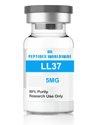
37 5 mg
Product Identity & Specifications
LL-37 is the sole human cathelicidin antimicrobial peptide, a 37-amino acid, α-helical, amphipathic peptide liberated from the C-terminal domain of the precursor protein hCAP-18 (human cationic antimicrobial protein, 18 kDa) by proteolytic processing. As the primary antimicrobial peptide produced by human neutrophils, epithelial cells, and keratinocytes, LL-37 occupies a central position in innate immune defense at mucosal surfaces and barrier tissues. Beyond its direct microbicidal activity, LL-37 functions as a master immunomodulator — recruiting immune cells to sites of infection, modulating Toll-like receptor (TLR) responses, promoting wound healing and angiogenesis, and neutralizing bacterial endotoxins. The 5 mg format is optimized for antimicrobial susceptibility assays, membrane permeabilization studies, immunomodulation research, and host defense peptide structure-function analysis.
| Specification | Detail |
|---|---|
| Peptide Name | LL-37 (hCAP-18 134-170, human cathelicidin) |
| Amino Acid Sequence | Leu-Leu-Gly-Asp-Phe-Phe-Arg-Lys-Ser-Lys-Glu-Lys-Ile-Gly-Lys-Glu-Phe-Lys-Arg-Ile-Val-Gln-Arg-Ile-Lys-Asp-Phe-Leu-Arg-Asn-Leu-Val-Pro-Arg-Thr-Glu-Ser |
| Sequence (Single Letter) | LLGDFFRKSKEKIGKEFKRIVQRIKDFLRNLVPRTES |
| CAS Registry Number | 154947-66-7 (hCAP-18 precursor) |
| Molecular Formula | C₂₀₅H₃₄₀N₆₀O₅₃ |
| Molecular Weight | 4493.31 g/mol (monoisotopic); 4496.33 g/mol (average) |
| Number of Amino Acids | 37 |
| Net Charge (pH 7) | +6 |
| Secondary Structure | α-Helical (in membrane-mimetic environments) |
| Purity (HPLC) | ≥98% |
| Physical Appearance | White to off-white lyophilized powder |
| Solubility | ≥1 mg/mL in sterile water; limited solubility in PBS/saline |
| Storage Condition | -20°C, desiccated, protected from light |
| Shelf Life (Lyophilized) | 24 months at recommended storage |
| Research Use Classification | Research Use Only (RUO) — Not for human or veterinary use |
Reviewed by: HKPEPTIDE WORLDWIDE Research Team | Last Updated: August 08, 2026
Research Background
The Cathelicidin Family: Evolution of Mammalian Host Defense Peptides
Antimicrobial peptides (AMPs) represent one of the most ancient and conserved elements of innate immunity, found in organisms ranging from plants and invertebrates to mammals. In humans, two major families of AMPs are recognized: the defensins (α-defensins and β-defensins) and the cathelicidins. While multiple defensin genes exist in the human genome, a striking evolutionary feature is that humans possess only a single cathelicidin gene — CAMP (cathelicidin antimicrobial peptide) — in contrast to cattle and pigs, which encode numerous cathelicidin variants. The human CAMP gene, located on chromosome 3p21.3, encodes the 170-amino acid preproprotein hCAP-18, which comprises an N-terminal signal peptide, a highly conserved cathelin-like prodomain (homologous to the cysteine protease inhibitor cathelin), and the C-terminal 37-residue antimicrobial domain — LL-37. The evolutionary conservation of a single cathelicidin gene in humans suggests that LL-37 serves non-redundant and essential roles in human host defense that cannot be compensated by other AMP families (Zanetti, 2004; Dürr et al., 2006).
Proteolytic Activation: From Proprotein to Active Peptide
LL-37 is stored as the inactive proform hCAP-18 in the specific granules of neutrophils and is secreted into the extracellular milieu upon neutrophil degranulation at sites of infection or inflammation. Proteolytic processing — the cleavage of the cathelin prodomain from the C-terminal LL-37 domain — is executed extracellularly by host serine proteases. The principal activating enzymes include kallikrein 5 and kallikrein 7 in the skin (cleaving after alanine and phenylalanine residues, respectively), proteinase 3 from neutrophils (cleaving after alanine), and gastricsin (pepsinogen C) in the gastric mucosa (cleaving after leucine). Additionally, matrix metalloproteinases (MMPs) produced by keratinocytes, including MMP-9, can process hCAP-18. This requirement for extracellular proteolytic activation provides a spatial and temporal regulatory mechanism, ensuring that the potent cytolytic activity of LL-37 is confined to sites where microbial challenge and host defense are co-localized (Sørensen et al., 2001; Yamasaki et al., 2006).
Structural Biology: The Amphipathic Helix Paradigm
Structural investigations of LL-37 by circular dichroism (CD) spectroscopy and nuclear magnetic resonance (NMR) have established that the peptide adopts a predominantly random coil conformation in aqueous solution but undergoes a dramatic conformational transition to an α-helical structure upon interaction with lipid membranes or in membrane-mimetic solvents (SDS micelles, lipid bicelles). The helical structure spans approximately residues 2-31, with the N-terminal region (residues 2-12) being more hydrophobic and the C-terminal region (residues 13-31) more hydrophilic — a hallmark amphipathic helix architecture. The cationic nature (net charge +6 at neutral pH, contributed by 6 lysine and 5 arginine residues versus 3 aspartic acid and 1 glutamic acid residues) and the spatial segregation of hydrophobic and cationic residues on opposite faces of the helix underpin LL-37’s ability to selectively target negatively charged bacterial membranes over zwitterionic eukaryotic membranes. The flexible C-terminal tail (residues 32-37) is disordered even in membrane-bound states and may facilitate peptide oligomerization during pore formation (Wang, 2008; Oren et al., 1999; Porcelli et al., 2008).
LL-37 in Contemporary Innate Immunity and Host Defense Research
LL-37 has emerged as a model archetype for understanding the dual antimicrobial-immunomodulatory functionality that characterizes host defense peptides. Unlike conventional antibiotics that act through a single defined molecular target, LL-37 employs a multi-hit strategy — combining direct membrane disruption with modulation of host immune signaling pathways — that makes the development of bacterial resistance substantially more difficult. This multi-target mechanism has generated intense interest in the context of the global antimicrobial resistance (AMR) crisis, with LL-37 serving as a structural template for the rational design of synthetic antimicrobial peptidomimetics. Furthermore, the recognition that LL-37 deficiency or dysfunction is associated with increased susceptibility to infections in conditions such as atopic dermatitis, Kostmann syndrome (severe congenital neutropenia), and cystic fibrosis has driven research into LL-37 replacement and augmentation strategies (Nijnik & Hancock, 2009; Vandamme et al., 2012; Mookherjee et al., 2020).
Molecular Mechanisms
Mechanism 1: Membrane Targeting and Toroidal Pore Formation
The direct antimicrobial activity of LL-37 is mediated through a multi-step membrane disruption process that begins with electrostatic attraction between the cationic peptide (+6 net charge) and the anionic surfaces of bacterial membranes, which are enriched in negatively charged phospholipids (phosphatidylglycerol, cardiolipin) and decorated with teichoic acids (Gram-positive) or lipopolysaccharide (Gram-negative). This initial electrostatic binding is followed by peptide insertion into the lipid bilayer, driven by the hydrophobic face of the amphipathic helix. At a critical peptide-to-lipid ratio (P/L*), the accumulated peptide molecules induce a positive curvature strain in the membrane, leading to the formation of toroidal (wormhole) pores — structures in which the lipid headgroups bend continuously from one membrane leaflet to the other, lined by both peptide and lipid molecules. These toroidal pores, typically 2-5 nm in diameter, compromise the permeability barrier, dissipate the transmembrane electrochemical gradient, and cause leakage of cytoplasmic contents, culminating in bacterial cell death. This mechanism is distinct from the barrel-stave model (in which peptides form a structured channel with a purely peptidic lining) and accounts for the detergent-like membrane-disruptive activity observed for LL-37 at higher concentrations (Henzler-Wildman et al., 2004; Lee et al., 2011; Xhindoli et al., 2016).
Mechanism 2: Immunomodulation via Formyl Peptide Receptor-Like 1 (FPRL1) and P2X7
Beyond direct killing, LL-37 profoundly modulates the host immune response through receptor-mediated signaling. LL-37 is a potent chemoattractant for human neutrophils, monocytes, and CD4+ T-lymphocytes, with this chemotactic activity mediated through formyl peptide receptor-like 1 (FPRL1, also known as FPR2/ALX). FPRL1 engagement by LL-37 triggers the Gαi-mediated signaling cascade, resulting in MAPK/ERK activation, calcium mobilization, and cell migration. Additionally, LL-37 interacts with the purinergic receptor P2X7 on macrophages, modulating ATP-induced IL-1β processing and release. At low (sub-micromolar) concentrations, LL-37 suppresses TLR4-mediated inflammatory cytokine production (TNF-α, IL-6) through an incompletely characterized mechanism that may involve interference with LPS-TLR4-MD-2 complex formation or disruption of downstream MyD88-dependent signaling. This anti-endotoxin activity is physiologically significant, as LL-37 can neutralize the pro-inflammatory effects of LPS released from dying Gram-negative bacteria, preventing the excessive inflammation that can lead to septic shock (De Yang et al., 2000; Elssner et al., 2004; Mookherjee et al., 2006).
Mechanism 3: Wound Healing, Angiogenesis, and Re-Epithelialization
LL-37 is a key molecular participant in the cutaneous wound healing response. It is upregulated in keratinocytes at wound edges within hours of injury and contributes to all phases of wound repair. LL-37 promotes re-epithelialization by stimulating keratinocyte proliferation and migration through transactivation of the epidermal growth factor receptor (EGFR) via metalloproteinase-mediated release of membrane-bound EGFR ligands. Additionally, LL-37 exerts pro-angiogenic effects — stimulating endothelial cell proliferation, migration, and tube formation — through FPRL1-mediated signaling and VEGF (vascular endothelial growth factor) induction. In dermal fibroblasts, LL-37 upregulates the expression of extracellular matrix components (collagen types I and III, fibronectin) and modulates the activity of matrix metalloproteinases, promoting the transition from the inflammatory to the proliferative phase of wound healing. These pleiotropic wound-repair activities underscore LL-37’s role as a bridging molecule that coordinates innate immunity with tissue regeneration (Heilborn et al., 2003; Koczulla et al., 2003; Tokumaru et al., 2005; Carretero et al., 2008).
Mechanism 4: Modulation of Apoptosis and Programmed Cell Death Pathways
LL-37 exhibits a dual capacity to either promote or suppress apoptosis depending on cellular context and concentration — a property that has significant implications for infection resolution and cancer biology. At high micromolar concentrations, LL-37 induces apoptosis in bacteria-infected macrophages and neutrophils, potentially facilitating the clearance of infected cells and limiting pathogen dissemination. This pro-apoptotic effect is associated with mitochondrial outer membrane permeabilization (MOMP), cytochrome c release, and caspase-3 activation. Conversely, at lower concentrations, LL-37 can suppress apoptosis in primary endothelial cells and keratinocytes exposed to pro-apoptotic stimuli (serum deprivation, TNF-α/cycloheximide) through PI3K/Akt pathway activation and upregulation of anti-apoptotic Bcl-2 family members. In cancer research, LL-37 has been investigated for selective cytotoxicity toward certain tumor cell lines (oral squamous cell carcinoma, colon carcinoma), with proposed mechanisms involving mitochondrial membrane targeting and activation of the intrinsic apoptotic pathway (Barlow et al., 2006; Ren et al., 2013; Kuroda et al., 2015).
Research Applications
LL-37 5 mg is deployed in a variety of specialized research contexts:
-
Antimicrobial Susceptibility Testing (AST): Minimum inhibitory concentration (MIC) and minimum bactericidal concentration (MBC) determinations against Gram-positive (S. aureus, MRSA, S. epidermidis), Gram-negative (E. coli, P. aeruginosa, K. pneumoniae, A. baumannii), and fungal (C. albicans, C. neoformans) pathogens under varying ionic strength, pH, and medium conditions.
-
Membrane Permeabilization and Biophysical Studies: Liposome leakage assays (calcein, ANTS/DPX), surface plasmon resonance (SPR) for peptide-lipid binding kinetics, and solid-state NMR or CD spectroscopy to characterize membrane-bound peptide topology.
-
Neutrophil Extracellular Trap (NET) Research: Investigation of LL-37 as a NET component and its role in NET-mediated bacterial killing, including quantification of NET-associated LL-37 by immunofluorescence and evaluation of NET-LL-37 bactericidal activity.
-
Biofilm Disruption and Anti-Biofilm Activity: Studies of LL-37 effects on established biofilms (crystal violet staining, confocal microscopy with LIVE/DEAD staining), and assessment of LL-37 synergy with conventional antibiotics in biofilm eradication.
-
Immunomodulation and Cytokine Profiling: LPS-stimulated PBMC or macrophage cultures (THP-1, RAW 264.7) examining LL-37 effects on cytokine/chemokine secretion (multiplex bead array: TNF-α, IL-1β, IL-6, IL-8, IL-10, MCP-1).
-
Wound Healing and Epithelial Biology: Scratch wound migration assays, keratinocyte proliferation (BrdU/MTS), and in-vitro angiogenesis assays (endothelial tube formation on Matrigel) to evaluate LL-37’s role in tissue repair.
Quality Control & Analytical Specifications
| Analytical Method | Specification | Acceptance Criteria |
|---|---|---|
| RP-HPLC Purity | C18, 214 nm UV detection | ≥98.0% peak area |
| ESI-TOF Mass Spectrometry | Positive ion mode | ⁴⁺ 1124.8 ± 0.5 Da; ⁵⁺ 900.3 ± 0.5 Da |
| Amino Acid Analysis | Post-column ninhydrin detection | ±10% of theoretical composition |
| Peptide Content (Net Peptide) | Elemental analysis (N%) | ≥80% |
| Residual TFA | Ion chromatography | ≤1.0% |
| Water Content (Karl Fischer) | Coulometric titration | ≤8.0% w/w |
| Endotoxin | LAL kinetic chromogenic | ≤1.0 EU/mg |
| Appearance | Visual inspection | White to off-white lyophilized powder |
| Solubility | 1 mg/mL in sterile H₂O | Clear, colorless solution |
| Secondary Structure | CD spectroscopy (220 nm ellipticity) | α-Helical content ≥40% in 50% TFE |
| Antimicrobial Activity | MIC S. aureus ATCC 25923 | 8-32 μg/mL (reference range) |
Available Configurations
| Product Variant | Catalog Number | Quantity | Format |
|---|---|---|---|
| LL-37 5 mg (current page) | LL37-005-USA | 5 mg per vial | Individual vial |
| Custom Bulk Orders | LL37-BULK-USA | Inquire | Custom quantities for institutional procurement |
Frequently Asked Questions (FAQ)
1. What is LL-37 and how is it derived from cathelicidin?
LL-37 is the biologically active C-terminal antimicrobial peptide (residues 134-170) liberated from the human cathelicidin precursor protein hCAP-18 (human cationic antimicrobial protein, 18 kDa). The CAMP gene on chromosome 3p21.3 encodes the full-length preproprotein, which consists of an N-terminal signal peptide (residues 1-30), a highly conserved cathelin-like prodomain (residues 31-130, containing four disulfide bonds and structurally homologous to cystatin protease inhibitors), and the C-terminal 37-amino acid antimicrobial domain (residues 131-170). The peptide was named LL-37 because it begins with two consecutive leucine residues (LL) and contains a total of 37 amino acids. Proteolytic processing — which removes the cathelin prodomain — is carried out extracellularly by host serine proteases including kallikrein 5 (in skin), proteinase 3 (in neutrophils), and gastricsin (in gastric mucosa). hCAP-18 is stored as the inactive proform in neutrophil-specific granules and is also constitutively expressed or inducibly expressed in epithelial cells of the skin, respiratory tract, gastrointestinal tract, and urogenital tract, positioned for immediate deployment upon microbial challenge.
2. What are the dual antimicrobial and immunomodulatory properties of LL-37?
LL-37 embodies a dual-function host defense paradigm. Its direct antimicrobial activity is mediated through electrostatic binding to anionic bacterial membrane surfaces, followed by insertion of its amphipathic α-helix, toroidal pore formation, and membrane permeabilization leading to cell death. This mechanism operates broadly against Gram-positive bacteria, Gram-negative bacteria, certain fungi, and enveloped viruses. Its immunomodulatory functions — which often occur at peptide concentrations below those required for direct killing — encompass a remarkably diverse set of activities: (a) chemotaxis of neutrophils, monocytes/macrophages, mast cells, and CD4+ T-lymphocytes to sites of infection; (b) modulation of TLR signaling, including suppression of TLR4-mediated TNF-α and IL-6 production in response to LPS (endotoxin neutralization); (c) stimulation of IL-8 secretion from epithelial cells and keratinocytes; (d) promotion of wound healing through EGFR transactivation, keratinocyte migration, and angiogenesis; (e) regulation of apoptosis in neutrophils and epithelial cells; (f) degranulation of mast cells with histamine release; (g) and modulation of dendritic cell differentiation and antigen presentation. This multifunctionality has earned LL-37 the designation “alarmin” — an endogenous danger signal that both alerts and calibrates the immune response.
3. How should LL-37 5 mg be stored and reconstituted?
Lyophilized LL-37 5 mg must be stored at -20°C in a desiccated, light-protected environment, achieving 24-month stability under these conditions. Reconstitution of LL-37 requires special attention due to its highly cationic and amphipathic nature. Sterile water for injection is the recommended reconstitution solvent; the peptide is soluble at ≥1 mg/mL with gentle vortexing and brief sonication (10-15 seconds). Importantly, LL-37 should NOT be initially reconstituted in phosphate-buffered saline (PBS) or physiological saline, as the high salt concentration and presence of divalent cations can induce peptide aggregation and precipitation. If PBS is required for the final experimental buffer, first reconstitute the peptide in sterile water at 10× the desired final concentration, then dilute into PBS. Use low-protein-binding polypropylene tubes for aliquoting. Upon reconstitution, prepare single-use aliquots and store at -20°C. Reconstituted LL-37 retains >90% antimicrobial activity for 14-21 days at -20°C. Avoid repeated freeze-thaw cycles. The peptide should be protected from oxidizing conditions and prolonged light exposure.
4. What is the antimicrobial spectrum of LL-37?
LL-37 exhibits broad-spectrum antimicrobial activity encompassing both Gram-positive and Gram-negative bacteria, as well as activity against certain fungi and enveloped viruses. Representative MIC (minimum inhibitory concentration) values in standard Mueller-Hinton broth include: Staphylococcus aureus (8-32 μg/mL), methicillin-resistant S. aureus / MRSA (16-64 μg/mL), Streptococcus pyogenes / Group A Streptococcus (4-16 μg/mL), Escherichia coli (8-32 μg/mL), Pseudomonas aeruginosa (16-64 μg/mL), Klebsiella pneumoniae (16-32 μg/mL), Acinetobacter baumannii (8-32 μg/mL), and Candida albicans (16-64 μg/mL). It is critical to note that these MIC values are highly sensitive to assay conditions — increasing salt concentration (NaCl, divalent cations), the presence of serum or plasma proteins, or the use of rich media can increase apparent MIC values substantially (2- to 8-fold elevation) due to electrostatic shielding, peptide sequestration, or proteolytic degradation. Researchers should carefully control and report buffer/media composition when conducting antimicrobial susceptibility assays with LL-37.
5. What purity level is verified for LL-37 5 mg?
HKPEPTIDE WORLDWIDE supplies LL-37 5 mg at ≥98% purity as verified by reverse-phase HPLC analysis using a C18 column with UV detection at 214 nm. Peptide identity and molecular weight are confirmed by electrospray ionization time-of-flight mass spectrometry (ESI-TOF MS). Due to the size and charge of LL-37 (4496 Da, +6 charge), multiply charged ion envelopes (³⁺ through ⁷⁺) are observed; the ⁴⁺ ion at m/z 1124.8 (±0.5 Da) and the ⁵⁺ ion at m/z 900.3 (±0.5 Da) are typically the most abundant charge states. Each batch includes a comprehensive Certificate of Analysis (CoA) documenting HPLC chromatogram, mass spectrum, peptide content (net peptide analysis via elemental nitrogen determination), residual trifluoroacetic acid (TFA), Karl Fischer water content, and LAL endotoxin level. Researchers may request batch-specific CoA documentation for regulatory compliance, grant reporting, or publication support.
References & Further Reading
-
Zanetti, M. (2004). Cathelicidins, multifunctional peptides of the innate immunity. Journal of Leukocyte Biology, 75(1), 39–48.
-
Dürr, U. H. N., Sudheendra, U. S., & Ramamoorthy, A. (2006). LL-37, the only human member of the cathelicidin family of antimicrobial peptides. Biochimica et Biophysica Acta (BBA) - Biomembranes, 1758(9), 1408–1425.
-
Sørensen, O. E., Follin, P., Johnsen, A. H., Calafat, J., Tjabringa, G. S., Hiemstra, P. S., & Borregaard, N. (2001). Human cathelicidin, hCAP-18, is processed to the antimicrobial peptide LL-37 by extracellular cleavage with proteinase 3. Blood, 97(12), 3951–3959.
-
Wang, G. (2008). Structures of human host defense cathelicidin LL-37 and its smallest antimicrobial peptide KR-12 in lipid micelles. Journal of Biological Chemistry, 283(47), 32637–32643.
-
De Yang, Chen, Q., Schmidt, A. P., Anderson, G. M., Wang, J. M., Wooters, J., Oppenheim, J. J., & Chertov, O. (2000). LL-37, the neutrophil granule- and epithelial cell-derived cathelicidin, utilizes formyl peptide receptor-like 1 (FPRL1) as a receptor to chemoattract human peripheral blood neutrophils, monocytes, and T cells. Journal of Experimental Medicine, 192(7), 1069–1074.
-
Heilborn, J. D., Nilsson, M. F., Kratz, G., Weber, G., Sørensen, O., Borregaard, N., & Ståhle-Bäckdahl, M. (2003). The cathelicidin anti-microbial peptide LL-37 is involved in re-epithelialization of human skin wounds and is lacking in chronic ulcer epithelium. Journal of Investigative Dermatology, 120(3), 379–389.
-
Koczulla, R., von Degenfeld, G., Kupatt, C., Krötz, F., Zahler, S., Gloe, T., Issbrücker, K., Unterberger, P., Zaiou, M., Lebherz, C., Karl, A., Raake, P., Pfosser, A., Boekstegers, P., Welsch, U., Hiemstra, P. S., Vogelmeier, C., Gallo, R. L., Clauss, M., & Bals, R. (2003). An angiogenic role for the human peptide antibiotic LL-37/hCAP-18. Journal of Clinical Investigation, 111(11), 1665–1672.
-
Mookherjee, N., Brown, K. L., Bowdish, D. M. E., Doria, S., Falsafi, R., Hokamp, K., Roche, F. M., Mu, R., Doho, G. H., Pistolic, J., Powers, J. P., Bryan, J., Brinkman, F. S. L., & Hancock, R. E. W. (2006). Modulation of the TLR-mediated inflammatory response by the endogenous human host defense peptide LL-37. Journal of Immunology, 176(4), 2455–2464.
-
Nijnik, A., & Hancock, R. E. W. (2009). The roles of cathelicidin LL-37 in immune defences and novel clinical applications. Current Opinion in Hematology, 16(1), 41–47.
-
Xhindoli, D., Pacor, S., Benincasa, M., Scocchi, M., Gennaro, R., & Tossi, A. (2016). The human cathelicidin LL-37 — A pore-forming antibacterial peptide and host-cell modulator. Biochimica et Biophysica Acta (BBA) - Biomembranes, 1858(3), 546–566.
-
Carretero, M., Escámez, M. J., García, M., Duarte, B., Holguín, A., Retamosa, L., Jorcano, J. L., Río, M. D., & Larcher, F. (2008). In vitro and in vivo wound healing-promoting activities of human cathelicidin LL-37. Journal of Investigative Dermatology, 128(1), 223–236.
-
Vandamme, D., Landuyt, B., Luyten, W., & Schoofs, L. (2012). A comprehensive summary of LL-37, the factotum human cathelicidin peptide. Cellular Immunology, 280(1), 22–35.
Related Research & Resources
| Resource | Description |
|---|---|
| Healing Regenerative Peptides Hub | Complete research overview & methodology hub |
| Ll 37 5 Mg Cathelicidin Antimicrobial Pe | Related research peptide product |
| Ll 37 5 Mg Research Peptide Usa | Related research peptide product |
| Understanding Safety Discussions Around Peptides | Latest research insights & methodology |
View Complete Research Guide »
Compliance Statement
This LL-37 5 mg research peptide is supplied exclusively as a Research Use Only (RUO) product for in-vitro laboratory investigations and preclinical scientific research conducted within qualified research facilities. It is not manufactured in accordance with current Good Manufacturing Practice (cGMP) regulations and is not intended, approved, or labeled for human administration, veterinary therapeutic use, clinical diagnosis, treatment of any disease or medical condition, or any form of therapeutic application whatsoever. Researchers bear sole responsibility for ensuring compliance with all applicable federal, state, and institutional regulations governing laboratory chemical and peptide handling, storage, use, and disposal. Any research protocol involving this product must receive appropriate institutional oversight, including IACUC approval for in-vivo research. HKPEPTIDE WORLDWIDE expressly disclaims any liability arising from improper use, misuse, diversion, or unauthorized application of this compound. By purchasing this product, the researcher acknowledges and agrees to these terms of use.
For Certificate of Analysis requests, technical inquiries, or institutional procurement, contact the HKPEPTIDE WORLDWIDE Research Support Team.