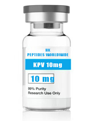
kpv 10 mg
Product Identity & Specifications
KPV is a minimalist tripeptide consisting of lysine (Lys/K), proline (Pro/P), and valine (Val/V) — the three C-terminal amino acid residues of alpha-melanocyte-stimulating hormone (α-MSH). Derived from the post-translational processing of proopiomelanocortin (POMC), α-MSH is a 13-amino acid neuropeptide with well-characterized roles in pigmentation, energy homeostasis, and immunomodulation. Structure-activity relationship (SAR) studies demonstrated that the C-terminal tripeptide Lys-Pro-Val retains the full anti-inflammatory potency of the parent tridecapeptide while completely lacking melanogenic activity, thereby representing a minimal pharmacophore for the anti-inflammatory actions of melanocortin peptides. The 10 mg format is optimized for extended in-vitro inflammatory pathway research, dose-response characterization, and multi-well experimental protocols requiring substantial material quantities.
| Specification | Detail |
|---|---|
| Peptide Name | KPV (α-MSH 11-13, Lys-Pro-Val) |
| Amino Acid Sequence | Lys-Pro-Val |
| Sequence (Single Letter) | KPV |
| CAS Registry Number | 67727-97-3 (α-MSH); KPV fragment — 13209-39-3 |
| Molecular Formula | C₁₆H₃₀N₄O₄ |
| Molecular Weight | 342.44 g/mol |
| Number of Amino Acids | 3 |
| Purity (HPLC) | ≥98% |
| Physical Appearance | White to off-white lyophilized powder |
| Solubility | ≥10 mg/mL in H₂O, PBS, and 0.9% saline |
| Storage Condition | -20°C, desiccated, protected from light |
| Shelf Life (Lyophilized) | 24 months at recommended storage |
| Research Use Classification | Research Use Only (RUO) — Not for human or veterinary use |
Reviewed by: HKPEPTIDE WORLDWIDE Research Team | Last Updated: August 08, 2026
Research Background
From Proopiomelanocortin Processing to the Minimal Anti-Inflammatory Pharmacophore
The proopiomelanocortin (POMC) gene encodes a 241-amino acid precursor polypeptide that undergoes tissue-specific post-translational processing by prohormone convertases (PC1/3 and PC2) to yield a family of biologically active peptides including adrenocorticotropic hormone (ACTH), the three melanocyte-stimulating hormones (α-MSH, β-MSH, γ-MSH), and β-endorphin. α-MSH, a tridecapeptide (Ser-Tyr-Ser-Met-Glu-His-Phe-Arg-Trp-Gly-Lys-Pro-Val-NH₂), was originally characterized for its melanogenic activity — the stimulation of eumelanin synthesis in melanocytes via the melanocortin 1 receptor (MC1R). However, the recognition that α-MSH also exerts profound anti-inflammatory and immunomodulatory effects, independent of its pigmentary actions, motivated systematic fragment-screening studies to identify the minimal sequence responsible for these anti-inflammatory properties (Luger et al., 1997; Catania et al., 2004).
Structure-Activity Relationship Studies: C-Terminal Domain Is Sufficient for Anti-Inflammatory Activity
Pioneering studies by Lipton, Catania, and colleagues in the 1980s and 1990s demonstrated that the C-terminal tripeptide of α-MSH — Lys-Pro-Val (KPV) — retains the capacity to suppress acute and chronic inflammatory responses in both in-vitro and in-vivo models. Systematic N-terminal and C-terminal truncation analysis revealed that while the core melanocortin signaling tetrapeptide His-Phe-Arg-Trp (the “message sequence”) is responsible for MC1R-mediated melanogenesis and cAMP accumulation, the C-terminal KPV tripeptide independently mediates anti-inflammatory effects without stimulating melanin production. This functional dissociation — melanogenic versus anti-inflammatory — established KPV as a valuable molecular probe for dissecting the signaling pathways underlying melanocortin-mediated immunomodulation (Hiltz & Lipton, 1989; Catania et al., 1996; Luger et al., 1998).
Discovery of Peripheral Anti-Inflammatory Action
A critical finding that catalyzed research interest in KPV was the demonstration that its anti-inflammatory effects are not restricted to the central nervous system (where α-MSH is predominantly expressed in the arcuate nucleus of the hypothalamus) but extend to peripheral tissues and inflammatory sites. KPV administered locally or systemically in rodent models of inflammation — including carrageenan-induced paw edema, picryl chloride-induced contact hypersensitivity, and dextran sulfate sodium (DSS)-induced colitis — produced significant reductions in inflammatory indices including edema volume, leukocyte infiltration, and pro-inflammatory cytokine levels (TNF-α, IL-1β, IL-6). These findings positioned KPV as a versatile anti-inflammatory research tool applicable across diverse tissue contexts (Luger et al., 1999; Getting et al., 1999).
KPV in Contemporary Inflammation and Immunology Research
Contemporary research interest in KPV has expanded significantly, driven by several converging factors: (1) the recognition that chronic low-grade inflammation (“inflammaging”) underlies numerous age-related pathologies; (2) the need for molecular tools that modulate inflammation without global immunosuppression; and (3) growing interest in melanocortin receptor signaling bias as a pharmacological strategy. KPV, with its selective anti-inflammatory profile and absence of melanogenic activity, serves as an ideal positive control and mechanistic probe in studies of MC1R/MC3R signaling bias, inflammasome regulation, and cytokine network modulation. Recent investigations have explored KPV-loaded nanoparticle formulations for targeted delivery to inflamed intestinal mucosa, opening new avenues for site-specific anti-inflammatory research (Xiao et al., 2016; Brzoska et al., 2008; Váradi et al., 2017).
Molecular Mechanisms
Mechanism 1: Melanocortin Receptor Engagement and cAMP/PKA Signaling
KPV exerts its anti-inflammatory effects through engagement with melanocortin receptors, with predominant activity at MC1R and MC3R subtypes. MC1R is expressed on macrophages, monocytes, neutrophils, dendritic cells, endothelial cells, and epithelial cells — a broad expression profile that mirrors KPV’s wide-ranging anti-inflammatory effects across multiple tissue types. Receptor engagement by KPV activates the stimulatory G-protein (Gsα)-adenylyl cyclase-cAMP-protein kinase A (PKA) signaling axis, leading to PKA-mediated phosphorylation of the transcription factor CREB (cAMP response element-binding protein). Phosphorylated CREB competes with NF-κB for the transcriptional coactivator CBP/p300, thereby indirectly suppressing NF-κB-dependent gene transcription. This mechanism — termed “transcriptional squelching” — represents a key pathway through which KPV attenuates pro-inflammatory gene expression without direct inhibition of NF-κB DNA-binding activity (Catania et al., 2004; Brzoska et al., 2008; Manna & Aggarwal, 1998).
Mechanism 2: NF-κB Pathway Inhibition and Cytokine Suppression
Beyond the indirect CREB-mediated mechanism, KPV directly interferes with NF-κB signaling at multiple nodes. Research in LPS-stimulated macrophages and TNF-α-activated endothelial cells has demonstrated that KPV treatment inhibits IκBα phosphorylation and degradation, thereby preventing NF-κB (p65/p50) nuclear translocation. This blockade of the canonical NF-κB activation pathway results in broad-spectrum suppression of NF-κB target genes including TNF-α, IL-1β, IL-6, IL-8 (CXCL8), MCP-1 (CCL2), ICAM-1, VCAM-1, and E-selectin. The coordinate downregulation of both pro-inflammatory cytokines and adhesion molecules distinguishes KPV from single-cytokine neutralizing strategies and reflects the multi-target nature of NF-κB pathway inhibition (Luger et al., 1999; Getting et al., 1999; Ichiyama et al., 1999).
Mechanism 3: NLRP3 Inflammasome Modulation
A more recently characterized mechanism of KPV action involves modulation of the NLRP3 (NOD-, LRR- and pyrin domain-containing protein 3) inflammasome — a multiprotein complex that serves as a critical sensor of cellular stress and microbial danger signals. NLRP3 inflammasome activation triggers caspase-1-mediated proteolytic maturation and secretion of the potent pro-inflammatory cytokines IL-1β and IL-18, as well as gasdermin D-mediated pyroptotic cell death. KPV has been shown to attenuate NLRP3 inflammasome activation in macrophages by reducing mitochondrial reactive oxygen species (mtROS) production and inhibiting the ASC (apoptosis-associated speck-like protein containing a CARD) oligomerization step required for inflammasome assembly. This mechanism is particularly relevant in research models of intestinal inflammation, where NLRP3 dysregulation contributes to disease pathogenesis (Deng et al., 2021; Xiao et al., 2016).
Mechanism 4: Epithelial Barrier Protection and Mucosal Homeostasis
In intestinal epithelial research, KPV demonstrates a unique capacity to preserve and restore epithelial barrier integrity under inflammatory conditions. Studies utilizing Caco-2 and T84 intestinal epithelial monolayers have shown that KPV treatment attenuates the TNF-α- and IFN-γ-induced decrease in transepithelial electrical resistance (TEER) — a quantitative measure of paracellular barrier function. Mechanistically, this barrier-protective effect is associated with preservation of tight junction protein expression and localization, including occludin, claudin-1, and ZO-1 (zonula occludens-1). Additionally, KPV reduces epithelial apoptosis in inflammatory conditions, as evidenced by decreased caspase-3 cleavage and annexin V positivity. This dual action — anti-inflammatory signaling combined with direct epithelial cytoprotection — makes KPV a valuable tool for studying the epithelial-immune interface in mucosal inflammation research (Dalmasso et al., 2008; Kannengiesser et al., 2008).
Research Applications
KPV 10 mg is deployed in a variety of specialized research contexts:
-
Inflammatory Bowel Disease (IBD) Modeling: Investigation of KPV in DSS-induced and TNBS-induced colitis models in rodents, including assessment of disease activity index (DAI), colon length, histological scoring, myeloperoxidase (MPO) activity, and cytokine profiling (TNF-α, IL-1β, IL-6, IL-10).
-
Macrophage Polarization and Inflammatory Signaling: Studies in LPS/IFN-γ-stimulated M1 macrophages and IL-4/IL-13-treated M2 macrophages examining KPV effects on polarization markers (iNOS, CD86, CD206, arginase-1) and inflammatory mediator release.
-
Endothelial Inflammation and Adhesion Molecule Research: TNF-α-stimulated HUVEC and HAEC cultures for quantification of ICAM-1, VCAM-1, and E-selectin surface expression (flow cytometry), leukocyte adhesion assays, and NF-κB reporter gene studies.
-
Nanoparticle and Drug Delivery System Development: KPV-loaded polymeric nanoparticles, liposomes, and hydrogel formulations for targeted delivery to inflamed mucosal surfaces, with characterization of encapsulation efficiency, release kinetics, and in-vitro bioactivity retention.
-
Skin Inflammation and Barrier Function: Assessment of KPV in UVB-irradiated keratinocyte cultures, contact hypersensitivity models, and dermal inflammation assays examining cytokine profiles, neutrophil infiltration, and epidermal barrier integrity.
-
Inflammasome Research: LPS/ATP and LPS/nigericin-stimulated macrophages for NLRP3 inflammasome activation studies, including caspase-1 activity, IL-1β secretion (ELISA), ASC speck formation (immunofluorescence), and mtROS measurement (MitoSOX).
Quality Control & Analytical Specifications
| Analytical Method | Specification | Acceptance Criteria |
|---|---|---|
| RP-HPLC Purity | C18, 214 nm UV detection | ≥98.0% peak area |
| ESI-TOF Mass Spectrometry | Positive ion mode | ⁺ 343.2 ± 0.5 Da |
| Amino Acid Analysis | Post-column ninhydrin detection | ±10% of theoretical composition (Lys:Pro:Val = 1:1:1) |
| Peptide Content (Net Peptide) | Elemental analysis (N%) | ≥80% |
| Residual TFA | Ion chromatography | ≤1.0% |
| Water Content (Karl Fischer) | Coulometric titration | ≤8.0% w/w |
| Endotoxin | LAL kinetic chromogenic | ≤1.0 EU/mg |
| Appearance | Visual inspection | White to off-white lyophilized powder |
| Solubility | 10 mg/mL in H₂O | Clear, colorless solution |
Available Configurations
| Product Variant | Catalog Number | Quantity | Format |
|---|---|---|---|
| KPV 10 mg (current page) | KPV-010-USA | 10 mg per vial | Individual vial |
| Custom Bulk Orders | KPV-BULK-USA | Inquire | Custom quantities for institutional procurement |
Frequently Asked Questions (FAQ)
1. What is KPV and what is its amino acid composition?
KPV is a tripeptide composed of lysine (Lys, K), proline (Pro, P), and valine (Val, V), representing the C-terminal fragment of alpha-melanocyte-stimulating hormone (α-MSH). These three amino acids — positions 11 through 13 of the full-length α-MSH tridecapeptide — were identified through systematic structure-activity relationship studies as the minimal sequence sufficient to retain the anti-inflammatory properties of the parent hormone. Despite consisting of only three amino acid residues and a molecular weight of just 342.44 g/mol, KPV exhibits potent anti-inflammatory activity across multiple tissue types and inflammatory models. The tripeptide is acetylated at the N-terminus and amidated at the C-terminus in its endogenous context (as part of α-MSH), though the synthetic non-acetylated, non-amidated form retains substantial bioactivity and is more commonly employed in research applications due to synthetic accessibility. The presence of proline in the central position imparts conformational constraint that may contribute to receptor recognition and proteolytic stability relative to unstructured tripeptides.
2. What is the mechanism of KPV’s anti-inflammatory activity?
KPV exerts its anti-inflammatory effects through a multi-pronged mechanistic framework. The primary pathway involves engagement with melanocortin receptors — predominantly MC1R and MC3R — which are broadly expressed on immune cells (macrophages, monocytes, neutrophils, dendritic cells), endothelial cells, and epithelial cells. Receptor activation stimulates Gsα-mediated adenylyl cyclase activity, elevating intracellular cAMP and activating PKA. PKA then phosphorylates CREB, and the resulting phospho-CREB-CBP/p300 complex competes with NF-κB for limiting pools of transcriptional coactivators, functionally squelching NF-κB-dependent gene transcription. Additionally, KPV directly inhibits the canonical NF-κB pathway by preventing IκBα phosphorylation and degradation, thereby blocking p65/p50 nuclear translocation. A third mechanism, elucidated more recently, involves suppression of the NLRP3 inflammasome — KPV reduces mitochondrial ROS production and attenuates ASC oligomerization, decreasing caspase-1 activation and subsequent IL-1β/IL-18 secretion. Collectively, these interconnected mechanisms produce coordinate suppression of multiple pro-inflammatory mediators including TNF-α, IL-1β, IL-6, IL-8, MCP-1, ICAM-1, and VCAM-1.
3. How should KPV 10 mg be stored in the laboratory?
Lyophilized KPV 10 mg must be stored at -20°C in a desiccated, light-protected environment, achieving 24-month stability under these conditions. For reconstitution, sterile water for injection, phosphate-buffered saline (PBS, pH 7.4), or 0.9% sterile saline is recommended. Due to its small size (tripeptide) and excellent aqueous solubility (≥10 mg/mL), KPV dissolves readily with gentle vortexing and may not require sonication. Upon reconstitution, prepare single-use or limited-use aliquots in sterile polypropylene or low-protein-binding tubes and store at -20°C. Reconstituted KPV retains >90% bioactivity for 21 days at -20°C. Avoid repeated freeze-thaw cycles. The peptide is relatively stable across pH 4.0-8.0 but should be protected from prolonged exposure to strongly alkaline conditions (pH > 8.5) that may promote deamidation or backbone hydrolysis. Researchers working with cell culture should sterile-filter (0.22 μm) reconstituted KPV before addition to culture media.
4. What types of research models utilize KPV?
KPV is employed across a broad spectrum of inflammation-focused research models. In in-vitro settings, common applications include LPS-stimulated RAW 264.7 and THP-1 macrophage cultures for cytokine profiling and NF-κB pathway analysis; TNF-α-activated HUVEC monolayers for adhesion molecule expression and leukocyte-endothelial interaction studies; and Caco-2/T84 intestinal epithelial barrier models for TEER measurements and tight junction protein analysis. In-vivo research applications are equally diverse, encompassing DSS-induced and TNBS-induced colitis models in mice and rats (with endpoints including body weight change, disease activity index, colon length, histopathology, and mucosal cytokine levels); carrageenan-induced paw edema and zymosan-induced peritonitis for acute inflammation assessment; contact hypersensitivity models (DNFB, oxazolone) for T-cell-mediated skin inflammation; and UVB-induced dermal inflammation models examining erythema, neutrophil infiltration, and COX-2 expression. The 10 mg vial format supports extended multi-group in-vivo protocols and comprehensive dose-response characterization.
5. What purity level is verified for KPV 10 mg?
HKPEPTIDE WORLDWIDE supplies KPV 10 mg at ≥98% purity as verified by reverse-phase HPLC analysis using a C18 column with UV detection at 214 nm. Peptide identity and molecular weight are confirmed by electrospray ionization time-of-flight mass spectrometry (ESI-TOF MS) with the expected ⁺ ion at m/z 343.2 (±0.5 Da). Each batch includes a comprehensive Certificate of Analysis (CoA) documenting HPLC chromatogram, mass spectrum, peptide content (net peptide analysis via elemental nitrogen determination), residual trifluoroacetic acid (TFA), Karl Fischer water content, and LAL endotoxin level. Researchers may request batch-specific CoA documentation for regulatory compliance, grant reporting, or publication support.
References & Further Reading
-
Catania, A., Gatti, S., Colombo, G., & Lipton, J. M. (2004). Targeting melanocortin receptors as a novel strategy to control inflammation. Pharmacological Reviews, 56(1), 1–29.
-
Luger, T. A., Scholzen, T., & Grabbe, S. (1997). The role of α-melanocyte-stimulating hormone in cutaneous biology. Journal of Investigative Dermatology, 2(2), 87–93.
-
Hiltz, M. E., & Lipton, J. M. (1989). Anti-inflammatory activity of a COOH-terminal fragment of the neuropeptide α-MSH. FASEB Journal, 3(11), 2282–2284.
-
Getting, S. J., Gibbs, L., Clark, A. J. L., Flower, R. J., & Perretti, M. (1999). POMC gene-derived peptides activate melanocortin type 3 receptor on murine macrophages, suppressing cytokine release and inhibiting neutrophil migration. Journal of Immunology, 162(12), 7446–7453.
-
Luger, T. A., Brzoska, T., Scholzen, T. E., Kalden, D. H., Sunderkötter, C., Armstrong, C., & Ansel, J. (1999). The role of α-MSH as a modulator of cutaneous inflammation. Annals of the New York Academy of Sciences, 885, 368–377.
-
Xiao, B., Zhang, Z., Viennois, E., Kang, Y., Zhang, M., Han, M. K., Chen, J., & Merlin, D. (2016). Combination therapy for ulcerative colitis: Orally targeted nanoparticles prevent mucosal damage and relieve inflammation. Theranostics, 6(12), 2250–2266.
-
Brzoska, T., Luger, T. A., Maaser, C., Abels, C., & Böhm, M. (2008). α-Melanocyte-stimulating hormone and related tripeptides: Biochemistry, anti-inflammatory and protective effects in vitro and in vivo, and future perspectives for the treatment of immune-mediated inflammatory diseases. Endocrine Reviews, 29(5), 581–602.
-
Manna, S. K., & Aggarwal, B. B. (1998). α-Melanocyte-stimulating hormone inhibits the nuclear transcription factor NF-κB activation induced by various inflammatory agents. Journal of Immunology, 161(6), 2873–2880.
-
Ichiyama, T., Sakai, T., Catania, A., Barsh, G. S., Furukawa, S., & Lipton, J. M. (1999). Inhibition of peripheral NF-κB activation by central and peripheral α-MSH. Journal of Neuroimmunology, 99(2), 211–217.
-
Dalmasso, G., Charrier-Hisamuddin, L., Nguyen, H. T., Yan, Y., Sitaraman, S., & Merlin, D. (2008). PepT1-mediated tripeptide KPV uptake reduces intestinal inflammation. Gastroenterology, 134(1), 166–178.
-
Váradi, J., Harazin, A., Fenyvesi, F., Réti-Nagy, K., Gogolák, P., Vámosi, G., Bácskay, I., Fehér, P., Ujhelyi, Z., Vasvári, G., Róka, E., Haines, D., Deli, M. A., & Vecsernyés, M. (2017). Alpha-melanocyte stimulating hormone protects against cytokine-induced barrier damage in Caco-2 intestinal epithelial monolayers. PLoS ONE, 12(8), e0183587.
-
Deng, Z., Ni, J., Wu, X., Wei, H., & Peng, J. (2021). KPV (Lys-Pro-Val) peptide attenuates NLRP3 inflammasome activation and protects against dextran sulfate sodium-induced colitis through SIRT1. European Journal of Pharmacology, 907, 174217.
Related Research & Resources
| Resource | Description |
|---|---|
| Healing Regenerative Peptides Hub | Complete research overview & methodology hub |
| Kpv 10 Mg Anti Inflammatory Research Pep | Related research peptide product |
| Kpv 10 Mg Laboratory Grade Compounds | Related research peptide product |
| Why Anti Aging Peptide Research Is Growing Rapidly | Latest research insights & methodology |
View Complete Research Guide »
Compliance Statement
This KPV 10 mg research peptide is supplied exclusively as a Research Use Only (RUO) product for in-vitro laboratory investigations and preclinical scientific research conducted within qualified research facilities. It is not manufactured in accordance with current Good Manufacturing Practice (cGMP) regulations and is not intended, approved, or labeled for human administration, veterinary therapeutic use, clinical diagnosis, treatment of any disease or medical condition, or any form of therapeutic application whatsoever. Researchers bear sole responsibility for ensuring compliance with all applicable federal, state, and institutional regulations governing laboratory chemical and peptide handling, storage, use, and disposal. Any research protocol involving this product must receive appropriate institutional oversight, including IACUC approval for in-vivo research. HKPEPTIDE WORLDWIDE expressly disclaims any liability arising from improper use, misuse, diversion, or unauthorized application of this compound. By purchasing this product, the researcher acknowledges and agrees to these terms of use.
For Certificate of Analysis requests, technical inquiries, or institutional procurement, contact the HKPEPTIDE WORLDWIDE Research Support Team.