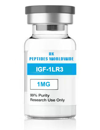
IGF-1 (Insulin-like Growth Factor-1 / Somato
Growth Factor ResearchReviewed by: HKPEPTIDE WORLDWIDE Research Team | Last Updated: 2026-08-08 | Document ID: HKPW-igf-1-research-grade-insulin-like-growth-factor-v1.0
1. Product Identity & Specifications
Insulin-like Growth Factor-1 (IGF-1) is a 70-amino acid single-chain polypeptide belonging to the insulin superfamily of growth factors, sharing approximately 48% sequence homology with proinsulin. HKPEPTIDE WORLDWIDE supplies recombinant human IGF-1 produced through validated expression systems, purified to ≥98% homogeneity, and lyophilized under controlled conditions to preserve the precise tertiary structure required for IGF-1R engagement. The product is available in two research configurations: 0.1 mg (100 µg) for pilot studies and receptor pharmacology, and 1 mg for extended experimental protocols.
IGF-1 is characterized by three intramolecular disulfide bonds—Cys⁶-Cys⁴⁸, Cys¹⁸-Cys⁶¹, and Cys⁴⁷-Cys⁵²—that constrain the polypeptide into a globular conformation with the A-domain and B-domain architecture characteristic of the insulin superfamily. The C-domain (residues 30–41) and D-domain (C-terminal extension absent in insulin) confer receptor subtype selectivity between IGF-1R and IR, making these structural elements critical for experimental interpretation.
| Parameter | Specification |
|---|---|
| Product Name | IGF-1 (Insulin-like Growth Factor-1 / Somatomedin C) |
| CAS Number | 67763-96-6 |
| Molecular Formula | C₃₃₁H₅₁₂N₉₄O₁₀₁S₇ |
| Molecular Weight | 7649 Da (average isotopic mass) |
| Amino Acid Length | 70 residues |
| Disulfide Bonds | Three intramolecular: Cys⁶-Cys⁴⁸, Cys¹⁸-Cys⁶¹, Cys⁴⁷-Cys⁵² |
| Available Configurations | 0.1 mg (100 µg) and 1.0 mg |
| Appearance | White to off-white lyophilized powder |
| Purity | ≥98% by RP-HPLC |
| Solubility | 10 mM HCl (initial reconstitution), then dilute in PBS or culture medium containing 0.1% BSA |
| Storage (Lyophilized) | -20°C, desiccated, protected from light |
| Storage (Reconstituted) | Aliquot in carrier protein-containing buffer; -20°C (≤3 months); avoid freeze-thaw |
| Product Grade | Research Use Only (RUO) |
| Peptide Content | ≥85% net peptide basis |
2. Research Background
IGF-1 occupies a central position in mammalian growth biology as the principal mediator of growth hormone (GH) action. The somatomedin hypothesis, first articulated by Salmon and Daughaday in 1957, proposed that GH stimulates skeletal growth indirectly through a circulating factor—subsequently identified as IGF-1. This discovery established the GH/IGF-1 axis as a fundamental endocrine signaling system governing somatic growth, cellular proliferation, differentiation, and metabolism across virtually every tissue.
The IGF-1 gene (located on chromosome 12q23.2 in humans) produces multiple mRNA transcripts through alternative splicing and promoter usage, yielding pro-peptides that undergo post-translational processing to produce the mature 70-amino acid peptide. Unlike insulin, which is stored in pancreatic β-cell secretory granules and released in response to glucose, IGF-1 is synthesized and secreted constitutively—primarily by the liver under GH stimulation—and circulates at nanomolar concentrations bound with high affinity to IGF-binding proteins (IGFBP-1 through -6). This IGFBP network creates a circulating reservoir that buffers IGF-1 bioavailability, extends its half-life from minutes to hours, and prevents the hypoglycemic episodes that would otherwise result from IGF-1’s inherent insulin-like activity.
The biological significance of IGF-1 has been demonstrated across diverse research domains. In skeletal muscle biology, IGF-1 drives myoblast proliferation and differentiation through the PI3K/Akt/mTOR axis, making it indispensable for studies of muscle hypertrophy, regeneration, and atrophy resistance. In bone biology, IGF-1 stimulates osteoblast proliferation, type I collagen synthesis, and longitudinal bone growth at the epiphyseal plate. In neurobiology, IGF-1 promotes neuronal survival, neurite outgrowth, and synaptic plasticity—effects that have positioned IGF-1R signaling as a target of interest in neurodegenerative disease research. In oncology research, dysregulation of the IGF-1/IGF-1R axis is implicated in tumor cell proliferation, survival, and resistance to chemotherapy, making IGF-1 an essential positive control for cancer cell line studies.
For the research community, HKPEPTIDE WORLDWIDE’s two-configuration approach—0.1 mg for assay development and 1 mg for comprehensive protocols—provides the flexibility needed for both pilot investigations and established experimental programs. At typical research concentrations of 1–100 ng/mL (0.13–13 nM), the 0.1 mg configuration provides sufficient material for approximately 1,000–100,000 assay wells; the 1 mg configuration supports approximately 10,000–1,000,000 wells.
3. Molecular Mechanisms
3.1 IGF-1 Receptor Binding and Activation
IGF-1 signals primarily through the type 1 IGF receptor (IGF-1R), a heterotetrameric (α₂β₂) receptor tyrosine kinase structurally homologous to the insulin receptor. IGF-1 binding to the extracellular α-subunits induces conformational changes that are transmitted to the intracellular β-subunit kinase domains, triggering trans-autophosphorylation of the tyrosine kinase activation loop (Tyr¹¹³¹, Tyr¹¹³⁵, Tyr¹¹³⁶). This initial phosphorylation event recruits insulin receptor substrate (IRS) proteins—primarily IRS-1 and IRS-2—through their phosphotyrosine-binding (PTB) and pleckstrin homology (PH) domains. IRS phosphorylation at multiple tyrosine residues creates docking sites for downstream SH2 domain-containing effectors, most critically the p85 regulatory subunit of PI3K.
IGF-1 binds IGF-1R with a Kd of approximately 0.1–0.5 nM, approximately 1,000-fold tighter than its binding to the insulin receptor (IR). Conversely, insulin binds IR with ~1,000-fold selectivity over IGF-1R. This exquisite selectivity arises from structural differences in the C-domain and D-domain: the IGF-1 C-domain (12 residues) is shorter than the insulin C-peptide (31 residues) and is not cleaved during processing, while the D-domain (8 residues at the C-terminus) is absent in insulin entirely. These domain differences determine which receptor is engaged at physiological concentrations.
3.2 PI3K/Akt/mTOR Pathway: The Hypertrophy Axis
The PI3K/Akt/mTOR signaling cascade constitutes the primary anabolic pathway downstream of IGF-1R. IGF-1 binding → IRS-1/2 phosphorylation → p85/p110 PI3K recruitment → PIP₃ generation at the plasma membrane → PDK1-mediated phosphorylation of Akt at Thr³⁰⁸ → mTORC2-mediated phosphorylation at Ser⁴⁷³ → full Akt activation. Active Akt then phosphorylates multiple substrates:
mTORC1 Activation: Akt phosphorylates and inactivates TSC2 (tuberin), relieving TSC1/TSC2-mediated inhibition of Rheb-GTP, which directly activates mTORC1. Active mTORC1 phosphorylates S6K1 (p70S6K) and 4E-BP1, driving ribosomal biogenesis and cap-dependent mRNA translation—the rate-limiting steps in protein synthesis and cellular hypertrophy.
FOXO Inactivation: Akt phosphorylates FOXO1 and FOXO3a at three conserved residues, promoting 14-3-3 binding and nuclear exclusion. This abrogates FOXO-dependent transcription of atrophy-related genes (Atrogin-1/MAFbx and MuRF1), thereby simultaneously promoting protein synthesis (mTORC1) and suppressing protein degradation (FOXO). This dual mechanism underlies IGF-1’s potent anti-atrophic effects in muscle wasting models.
GSK-3β Inhibition: Akt phosphorylates GSK-3β at Ser⁹, inactivating this kinase and relieving its inhibitory phosphorylation of glycogen synthase and eIF2B, thereby promoting glycogen synthesis and global translation initiation.
3.3 Ras/Raf/MEK/ERK Pathway: The Proliferation Axis
IRS-1/2 also recruit the Grb2/SOS complex through their multiple phosphotyrosine motifs, activating the Ras GTPase. Ras-GTP recruits Raf (MAPKKK) to the membrane, initiating the sequential phosphorylation cascade: Raf → MEK1/2 → ERK1/2. Active ERK translocates to the nucleus where it phosphorylates Elk-1, c-Fos, c-Jun, and other transcription factors regulating cyclin D1 expression and G1→S cell cycle progression. The ERK pathway primarily drives cellular proliferation, complementing the PI3K/Akt-mediated hypertrophic response—together producing the coordinated increase in cell size and cell number characteristic of tissue growth.
3.4 IGFBPs: Context-Dependent Bioavailability Regulation
IGF-1 bioavailability is modulated by six high-affinity IGF-binding proteins (IGFBP-1 through -6) and at least ten IGFBP-related proteins (IGFBP-rPs). In circulation, approximately 75% of IGF-1 exists in a ternary complex with IGFBP-3 (or IGFBP-5) and the acid-labile subunit (ALS), creating a 150-kDa complex that cannot cross the capillary endothelium. This ternary complex extends circulating half-life from ~10 minutes (free IGF-1) to ~15 hours. At the tissue level, IGFBP proteases (PAPP-A, PAPP-A2, MMPs) cleave IGFBPs, reducing their IGF-1 affinity and releasing free IGF-1 for receptor engagement. This proteolytic release mechanism provides spatially and temporally regulated IGF-1 bioavailability—a critical consideration for in vitro research where IGFBP supplementation or serum-containing media significantly influence experimental outcomes.
3.5 Cross-Talk with Integrin Signaling
In adherent cell types, IGF-1R signaling is functionally modulated by integrin-mediated adhesion. IGF-1 stimulates the tyrosine phosphorylation of focal adhesion kinase (FAK) and the adaptor protein p130Cas, linking IGF-1R to the actin cytoskeleton and promoting cell migration. This integrin/IGF-1R cross-talk is mediated by the formation of a ternary complex involving IGF-1R, αvβ3 integrin, and IRS-1, which localizes IGF-1 signaling to focal adhesions. In the absence of integrin engagement (e.g., cells in suspension), IGF-1 signaling through PI3K/Akt is preserved but ERK activation is attenuated, indicating matrix-dependence specifically for the proliferative arm of IGF-1 signaling. Researchers must account for this context-dependence when comparing IGF-1 effects across adherent versus suspension culture conditions.
4. Research Applications & Focus Areas
The 0.1 mg and 1 mg IGF-1 configurations are ideally suited for:
- Myogenesis and Muscle Hypertrophy Research: C2C12 and primary myoblast differentiation assays (0–7 day time courses), myotube diameter quantification, and fusion index analysis under IGF-1 treatment (10–100 ng/mL)
- Receptor Binding and Signaling Kinetics: Quantitative radioligand binding (¹²⁵I-IGF-1), phospho-Akt/phospho-ERK western blot time courses, and pathway-specific inhibitor studies (PI3K inhibitor LY294002, MEK inhibitor PD98059)
- Atrophy and Cachexia Models: Dexamethasone- or TNF-α-induced muscle atrophy in C2C12 myotubes with IGF-1 rescue; evaluation of MuRF1 and Atrogin-1/MAFbx expression by qPCR
- Bone and Cartilage Biology: Primary osteoblast proliferation and differentiation (ALP activity, mineralized nodule formation), chondrocyte matrix synthesis (³⁵S-sulfate incorporation), and growth plate explant studies
- Neuroprotection and Neurotrophic Factor Research: Primary neuronal cultures, SH-SY5Y differentiation, neurite outgrowth quantification, and PI3K/Akt-dependent neuroprotection against oxidative or excitotoxic stress
- Cancer Cell Line Studies: IGF-1R-dependent proliferation and survival signaling in MCF-7, MDA-MB-231, LNCaP, PC-3, and other IGF-1R-expressing tumor lines; combination studies with IGF-1R inhibitors (BMS-754807, linsitinib)
- Stem Cell Biology: IGF-1 supplementation of mesenchymal stem cell (MSC) and induced pluripotent stem cell (iPSC) differentiation protocols toward myogenic, osteogenic, and chondrogenic lineages
- Wound Healing and Tissue Regeneration: Keratinocyte and fibroblast proliferation and migration assays, collagen gel contraction models, and ex vivo skin organ culture with IGF-1 supplementation
- IGFBP Interaction Studies: SPR and ITC-based characterization of IGF-1/IGFBP binding kinetics; evaluation of IGFBP protease-mediated IGF-1 release
5. Quality Control & Analytical Specifications
| Test | Method | Acceptance Criteria |
|---|---|---|
| Purity | RP-HPLC (C18 column, 214 nm) | ≥98.0% |
| Molecular Weight | ESI-MS (intact + reduced/alkylated) | 7649 ± 1.0 Da |
| Disulfide Bond Integrity | LC-MS (non-reduced vs. DTT-reduced) | Correct three-disulfide pattern |
| Peptide Content | Amino Acid Analysis (AAA) | ≥85.0% |
| Sequence Confirmation | LC-MS/MS peptide mapping (trypsin + Glu-C) | ≥95% sequence coverage |
| Endotoxin | LAL Kinetic Chromogenic | ≤1.0 EU/mg |
| Water Content | Karl Fischer Titration | ≤5.0% |
| TFA Content | Ion Chromatography | ≤1.0% |
| Appearance | Visual Inspection (D65 illumination) | White to off-white powder |
| Biological Activity | IGF-1R phospho-Akt Ser⁴⁷³ assay (MCF-7, 10 ng/mL, 15 min) | ≥3-fold induction vs. unstimulated |
6. IGF-1 vs. Mechano Growth Factor (MGF): Comparative Analysis
IGF-1 and its splice variant MGF (mechano growth factor, IGF-1Ec in humans) share the first 49 amino acids but diverge in their C-terminal sequences due to alternative splicing. IGF-1 retains the full E-peptide-processed mature 70-aa peptide; MGF contains a unique 24-aa C-terminal extension that is not cleaved. Functionally, recombinant mature IGF-1 (supplied here) activates both myoblast proliferation (ERK-dependent) and differentiation/fusion (Akt-dependent) and is suitable for sustained signaling studies due to its stability. MGF preferentially activates the proliferation arm, does not promote differentiation, and has a shorter biological half-life. Researchers studying the early proliferative phase of muscle repair may prefer MGF; those studying sustained anabolic signaling or requiring stable receptor pharmacology should use mature IGF-1.
7. Tiered Wholesale Pricing
| Quantity | Price Per Vial (0.1 mg) | Price Per Vial (1 mg) | SKU |
|---|---|---|---|
| 1 Vial | $95.00 | $320.00 | HKPW-IGF1-01MG / HKPW-IGF1-1MG |
| 5 Vials | $85.50/vial ($427.50) | $288.00/vial ($1,440.00) | Multi-pack |
| 10 Vials | $76.00/vial ($760.00) | $256.00/vial ($2,560.00) | Multi-pack |
| 25+ Vials | Contact for bulk pricing | Contact for bulk pricing | HKPW-IGF1-BULK |
All prices in USD. Institutional and academic discounts available upon verification. Recombinant production costs vary by batch size; contact our team for custom synthesis volumes.
8. Frequently Asked Questions
Q: Why does IGF-1 require acidic conditions for initial reconstitution?
IGF-1 has an isoelectric point (pI) of approximately 8.2 and is poorly soluble at neutral pH in the absence of carrier proteins. Initial reconstitution in 10 mM HCl (pH ~2.0) ensures complete solubilization; subsequent dilution into PBS or culture medium containing 0.1–0.5% BSA prevents re-aggregation and surface adsorption, which can cause significant material loss at low concentrations.
Q: What is the role of the C-domain in receptor selectivity?
The IGF-1 C-domain (residues 30–41: Gly-Tyr-Gly-Ser-Ser-Ser-Arg-Arg-Ala-Pro-Gln-Thr) forms a surface-exposed loop that sterically hinders binding to the insulin receptor. Deletion of the C-domain increases IR cross-reactivity approximately 10-fold, while swapping the IGF-1 C-domain for the insulin C-peptide produces a hybrid with intermediate receptor selectivity. Researchers using IGF-1 analogs or IGF-1/insulin combinations must account for this structural basis of selectivity.
Q: How stable is reconstituted IGF-1?
IGF-1 is susceptible to aggregation, oxidation (Met⁵⁹), and deamidation at neutral-to-alkaline pH. Reconstituted IGF-1 in 10 mM HCl is stable for one week at 2–8°C. For cell culture applications, dilute working aliquots in PBS + 0.1% BSA, aliquot single-use volumes, and store at -20°C. Avoid repeated freeze-thaw cycles. Do not store in water alone—surface adsorption to plastic can exceed 50% at concentrations below 1 µg/mL.
Q: Can IGF-1 be used in serum-free culture conditions?
Yes, but with important caveats. Serum-free conditions eliminate IGFBP-mediated modulation of bioavailability. Supplementation with 0.1% BSA or 1 nM recombinant IGFBP-3 more accurately reflects physiological IGF-1 presentation and prevents rapid degradation by serum proteases. Researchers studying IGF-1R pharmacology should consider the effects of IGFBP presence or absence on dose-response relationships.
Q: What distinguishes recombinant IGF-1 from the LR3 analog?
IGF-1 LR3 contains an Arg substitution at Glu³ and a 13-aa N-terminal extension, conferring approximately 1,000-fold reduced affinity for IGFBPs while maintaining full IGF-1R agonism. LR3 is useful for experiments where IGFBP interference needs to be eliminated; however, its prolonged signaling kinetics (due to lack of IGFBP buffering) differ substantially from native IGF-1. HKPEPTIDE WORLDWIDE’s recombinant human IGF-1 represents the native sequence, providing physiologically relevant receptor pharmacology.
9. References & Further Reading
- Salmon WD Jr, Daughaday WH. A hormonally controlled serum factor which stimulates sulfate incorporation by cartilage in vitro. J Lab Clin Med. 1957. (The seminal somatomedin hypothesis paper — PMID: 13428999)
- LeRoith D, Werner H, Beitner-Johnson D, Roberts CT Jr. Molecular and cellular aspects of the insulin-like growth factor I receptor. Endocr Rev. 1995.
- Glass DJ. PI3 kinase regulation of skeletal muscle hypertrophy and atrophy. Curr Top Microbiol Immunol. 2010. (The foundational PI3K/Akt/mTOR/FOXO framework in muscle — PMID: 20020324)
- Batch-specific Certificate of Analysis (COA) included with every order
- Full HPLC chromatogram and ESI-MS spectrum available upon request
- Technical dossier provided with bulk wholesale orders
10. Internal Resources
- Research Peptide Quality Guide
- COA and Quality Standards
- Shipping & Handling Information
- Contact Our Research Team
11. Compliance Statement
FOR LABORATORY RESEARCH USE ONLY. This product is intended exclusively for in vitro laboratory research and scientific investigation. Not for human or veterinary diagnostic, therapeutic, or prophylactic use. Not a dietary supplement, pharmaceutical ingredient, or approved drug substance. IGF-1 is classified as a prohibited substance by the World Anti-Doping Agency (WADA) under category S2 (Peptide Hormones, Growth Factors, and Related Substances). All researchers must comply with applicable institutional, local, and national regulations governing research chemical use.
Related Research & Resources
| Resource | Description |
|---|---|
| Healing Regenerative Peptides Hub | Complete research overview & methodology hub |
| Igf 1 0 1 Mg 1 Mg Research Grade Insulin | Related research peptide product |
| Why Muscle Growth Peptide Research Is Expanding | Latest research insights & methodology |
View Complete Research Guide »
© 2026 HKPEPTIDE WORLDWIDE. All rights reserved. Document ID: HKPW-igf-1-research-grade-insulin-like-growth-factor-v1.0