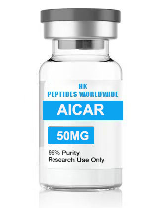
aicar 5 mg
Product Identity & Specifications
AICAR (5-Aminoimidazole-4-carboxamide ribonucleotide), also known as Acadesine or AICA ribonucleotide, is a cell-permeable adenosine analog and one of the most widely utilized pharmacological activators of AMP-activated protein kinase (AMPK) in biomedical research. The 5 mg format is optimized for precision laboratory experiments, pilot studies, and controlled in-vitro protocols where exact dosimetry and minimal material usage are critical.
| Specification | Detail |
|---|---|
| Full Chemical Name | 5-Aminoimidazole-4-carboxamide ribonucleotide |
| CAS Registry Number | 2627-69-2 |
| Molecular Formula | C₉H₁₅N₄O₈P |
| Molecular Weight | 338.21 g/mol |
| Synonyms | Acadesine, AICA Ribonucleotide, ZMP precursor |
| Purity (HPLC) | ≥98% |
| Physical Appearance | White to off-white lyophilized powder |
| Solubility | Water-soluble (≥10 mg/mL); soluble in PBS, DMSO |
| Storage Condition | -20°C, desiccated, protected from light |
| Shelf Life (Lyophilized) | 24 months at recommended storage |
| Research Use Classification | Research Use Only (RUO) — Not for human or veterinary use |
Reviewed by: HKPEPTIDE WORLDWIDE Research Team | Last Updated: August 08, 2026
Research Background
Discovery and Development of AICAR
AICAR was first synthesized and characterized in the mid-20th century as part of investigations into purine nucleotide biosynthesis. Its structural similarity to adenosine, coupled with the distinctive 5-aminoimidazole-4-carboxamide moiety, distinguished it from endogenous nucleosides and prompted extensive biochemical characterization. The compound was initially explored for its potential cardioprotective properties, receiving the investigational name “Acadesine” during clinical development for ischemia-reperfusion injury indications (Mullane, 1993; Mangano, 1997).
The AMPK Connection: A Paradigm Shift
The landmark discovery that fundamentally reshaped AICAR’s role in research occurred in the 1990s, when Corton and colleagues (1995) demonstrated that AICAR, following intracellular uptake and phosphorylation by adenosine kinase, is converted to 5-aminoimidazole-4-carboxamide ribonucleotide monophosphate (ZMP). ZMP functions as an AMP mimetic, binding to the γ-subunit of AMPK and triggering allosteric activation of the kinase. This finding established AICAR as the premier pharmacological tool for probing AMPK signaling — a discovery that catalyzed thousands of subsequent investigations into cellular energy homeostasis (Hardie et al., 1998).
AICAR in Contemporary Metabolic Research
In the decades since its AMPK-activating properties were elucidated, AICAR has become an indispensable research reagent in metabolic science. It is now routinely employed to investigate glucose transporter type 4 (GLUT4) translocation in skeletal muscle, hepatic gluconeogenesis suppression, fatty acid oxidation in adipose tissue, and mitochondrial biogenesis via PGC-1α signaling (Winder et al., 2000; Jørgensen et al., 2004). The compound’s ability to simulate exercise-like metabolic adaptations in sedentary models — including enhanced insulin sensitivity and oxidative capacity — has positioned AICAR centrally within exercise physiology and diabetes research (Narkar et al., 2008).
Molecular Mechanisms
Mechanism 1: Intracellular Phosphorylation to ZMP and AMPK Allosteric Activation
Upon entering the cell via nucleoside transporters, AICAR is rapidly phosphorylated by adenosine kinase to yield AICA ribonucleotide monophosphate, commonly designated ZMP. ZMP accumulates intracellularly and directly binds the cystathionine beta-synthase (CBS) domains within the γ-subunit of AMPK. The binding of ZMP to CBS domain 3 (site 3) induces a conformational change that promotes Thr172 phosphorylation on the α-subunit’s activation loop by upstream kinases — principally LKB1 (serine/threonine kinase 11) and Ca²⁺/calmodulin-dependent protein kinase kinase β (CaMKKβ). This dual mechanism — allosteric activation combined with enhanced phosphorylation — results in a 50- to 100-fold increase in AMPK catalytic activity (Hardie et al., 2012; Xiao et al., 2011).
Mechanism 2: AMPK-Mediated GLUT4 Translocation and Glucose Transport
Activated AMPK directly phosphorylates TBC1 domain family member 1 (TBC1D1) at Ser231 and related residues, triggering Rab-GTPase-dependent translocation of GLUT4-containing vesicles to the plasma membrane. In skeletal muscle and cardiac tissue, AICAR-stimulated AMPK activity increases glucose uptake by 2- to 5-fold in an insulin-independent manner. This mechanism is of particular research interest because it operates through a signaling cascade distinct from the canonical insulin/PI3K/Akt pathway, offering a parallel route to cellular glucose disposal that bypasses insulin resistance (Kurth-Kraczek et al., 1999; Treebak et al., 2006).
Mechanism 3: Acetyl-CoA Carboxylase (ACC) Phosphorylation and Fatty Acid Oxidation
AMPK activated by AICAR-derived ZMP phosphorylates and inactivates acetyl-CoA carboxylase (ACC) at Ser79 (ACC1) and Ser212 (ACC2). ACC inactivation reduces malonyl-CoA synthesis, relieving the allosteric inhibition of carnitine palmitoyltransferase I (CPT-1) — the rate-limiting enzyme for mitochondrial long-chain fatty acid import. The resultant increase in fatty acid β-oxidation has been quantified as a 1.5- to 3-fold elevation in palmitate oxidation rates across multiple tissue models. This mechanism constitutes a primary node through which AICAR shifts cellular metabolism from lipid storage toward oxidative disposal (Merrill et al., 1997; Winder & Hardie, 1996).
Mechanism 4: PGC-1α-Mediated Mitochondrial Biogenesis
Chronic AICAR treatment of cultured myotubes and in-vivo rodent models reproducibly upregulates peroxisome proliferator-activated receptor gamma coactivator 1-alpha (PGC-1α) expression via AMPK-dependent phosphorylation of histone deacetylase 5 (HDAC5) and subsequent activation of myocyte enhancer factor 2 (MEF2) transcription factors. PGC-1α coordinates mitochondrial DNA replication, respiratory chain subunit expression, and oxidative fiber-type switching. Research has demonstrated that 4 weeks of AICAR administration in murine models increases mitochondrial density by approximately 25-40% and enhances maximal oxygen consumption capacity (Zong et al., 2002; Jäger et al., 2007).
Research Applications
AICAR 5 mg is deployed across a broad spectrum of laboratory research disciplines:
-
AMPK Pathway Dissection: Pharmacological activation of AMPK to distinguish AMPK-dependent from AMPK-independent signaling events in kinase cascade analysis. Researchers employ AICAR alongside AMPK inhibitors (Compound C/dorsomorphin) and genetic models (AMPK knockout/knockdown) to establish mechanistic causality.
-
Glucose Homeostasis and Insulin Sensitivity Research: Investigation of insulin-independent GLUT4 translocation in L6 myotubes, C2C12 myoblasts, and primary human skeletal muscle cell cultures. Frequently used in comparative studies of type 2 diabetes pathobiology.
-
Hepatic Metabolism Studies: Examination of AICAR effects on gluconeogenic gene expression (PEPCK, G6Pase suppression), hepatic lipid droplet formation, and de novo lipogenesis in primary hepatocytes and HepG2 cell lines.
-
Cardiac Ischemia-Reperfusion Models: Preconditioning protocols in isolated perfused heart (Langendorff) preparations to study AMPK-mediated cardioprotective signaling, including infarct size limitation and contractile recovery.
-
Exercise Physiology and Muscle Biology: Research into endurance exercise mimetics, muscle fiber-type specification, and metabolic reprogramming in oxidative versus glycolytic muscle phenotypes.
-
Cancer Metabolism: Investigation of AMPK-mediated growth suppression, mTOR pathway inhibition, and metabolic vulnerability in tumor cell lines exhibiting deregulated energy sensing.
Quality Control & Analytical Specifications
Every batch of AICAR 5 mg supplied by HKPEPTIDE WORLDWIDE undergoes rigorous analytical characterization to ensure identity, purity, and consistency:
| Analytical Method | Specification | Acceptance Criteria |
|---|---|---|
| HPLC Purity | Reverse-phase C18, UV detection at 260 nm | ≥98.0% peak area |
| LC-MS Identity | Electrospray ionization, positive mode | Molecular ion ⁺ 339.1 ± 0.5 Da |
| Residual Solvents (GC-HS) | USP <467> | Class 3 solvents <0.5% |
| Water Content (Karl Fischer) | Coulometric titration | ≤5.0% w/w |
| Endotoxin | LAL kinetic chromogenic | ≤1.0 EU/mg |
| Appearance | Visual inspection | White to off-white powder |
| Reconstitution Clarity | 10 mg/mL in H₂O | Clear, colorless solution |
| Bioactivity (Cell-Based AMPK Phosphorylation Assay) | p-AMPKα (Thr172) ELISA in C2C12 cells | ≥3-fold increase vs. vehicle at 1 mM, 30 min |
Each shipment includes a batch-specific Certificate of Analysis (CoA) documenting all quality control parameters.
Available Configurations
| Product Variant | Catalog Number | Quantity | Format |
|---|---|---|---|
| AICAR 5 mg (current page) | AIC-005-USA | 5 mg per vial | Individual vial |
| AICAR 50 mg | AIC-050-USA | 50 mg per vial | Individual vial |
| Custom Bulk Orders | AIC-BULK-USA | Inquire | Custom quantities available upon request for institutional procurement |
Frequently Asked Questions (FAQ)
1. What is AICAR and how does it function in AMPK research?
AICAR (5-Aminoimidazole-4-carboxamide ribonucleotide) is a cell-permeable adenosine analog that, upon intracellular phosphorylation by adenosine kinase to ZMP (AICA ribonucleotide monophosphate), directly and allosterically activates AMP-activated protein kinase (AMPK). ZMP binds the γ-subunit CBS domains of AMPK, promoting phosphorylation at Thr172 by upstream kinases LKB1 and CaMKKβ. This activation precipitates downstream effects including GLUT4-mediated glucose uptake, fatty acid oxidation via ACC phosphorylation, and mitochondrial biogenesis through PGC-1α signaling. AICAR is the most widely employed pharmacological AMPK activator in biomedical research, with over 15,000 peer-reviewed publications referencing its use.
2. What concentration of AICAR is typically used in in-vitro studies?
In-vitro research protocols typically employ AICAR at concentrations ranging from 0.1 mM to 2 mM, with incubation periods varying from 30 minutes (for acute signaling readouts such as phospho-ACC or phospho-AMPK) to 24–48 hours (for transcriptional and metabolic reprogramming endpoints). Cell-type-specific optimization is recommended: primary hepatocytes often respond maximally at 0.5 mM, while C2C12 myotubes may require 1–2 mM for robust AMPK activation. The 5 mg vial provides sufficient material for approximately 75–150 assays at 1 mM in standard 6-well plate formats (2 mL working volume).
3. How should AICAR 5 mg be stored in a research laboratory?
Lyophilized AICAR 5 mg should be stored at -20°C in a desiccated, light-protected environment, under which conditions it remains stable for 24 months. Upon reconstitution in sterile, nuclease-free water, PBS (pH 7.4), or cell culture medium, aliquots should be prepared and stored at -20°C. Avoid repeated freeze-thaw cycles. Reconstituted solutions maintain ≥95% bioactivity for 30 days when stored at -20°C. Solubility exceeds 10 mg/mL in aqueous solvents. DMSO solutions (≥50 mg/mL) may be prepared for stock solution preparation, with subsequent dilution into aqueous media.
4. Is AICAR 5 mg suitable for in-vivo research models?
Yes, AICAR is extensively utilized in in-vivo rodent models. Published protocols employ intraperitoneal (IP) administration at doses of 0.25 to 1.0 g/kg body weight, typically dissolved in sterile saline. Chronic administration studies — lasting up to 8 weeks — have demonstrated consistent AMPK activation in skeletal muscle, liver, and adipose tissue. Researchers should design protocols with appropriate vehicle controls and consult species-specific pharmacokinetic data. Note that AICAR is not approved for human or veterinary therapeutic use and is supplied strictly for preclinical laboratory research.
5. What purity grade does HKPEPTIDE WORLDWIDE supply for AICAR 5 mg?
HKPEPTIDE WORLDWIDE supplies AICAR 5 mg at ≥98% purity verified by reverse-phase HPLC with UV detection at 260 nm. Identity confirmation is performed via LC-MS (ESI+, molecular ion ⁺ m/z 339.1). Each batch is accompanied by a comprehensive Certificate of Analysis (CoA) that includes retention time, purity percentage, mass spectrum, residual solvent analysis, water content, and endotoxin levels. Laboratory researchers can request batch-specific CoA documentation at any time.
References & Further Reading
-
Corton, J. M., Gillespie, J. G., Hawley, S. A., & Hardie, D. G. (1995). 5-Aminoimidazole-4-carboxamide ribonucleoside: A specific method for activating AMP-activated protein kinase in intact cells? European Journal of Biochemistry, 229(2), 558–565.
-
Hardie, D. G., Ross, F. A., & Hawley, S. A. (2012). AMPK: A nutrient and energy sensor that maintains energy homeostasis. Nature Reviews Molecular Cell Biology, 13(4), 251–262.
-
Merrill, G. F., Kurth, E. J., Hardie, D. G., & Winder, W. W. (1997). AICA riboside increases AMP-activated protein kinase, fatty acid oxidation, and glucose uptake in rat muscle. American Journal of Physiology, 273(6), E1107–E1112.
-
Winder, W. W., & Hardie, D. G. (1996). Inactivation of acetyl-CoA carboxylase and activation of AMP-activated protein kinase in muscle during exercise. American Journal of Physiology, 270(2), E299–E304.
-
Narkar, V. A., Downes, M., Yu, R. T., Embler, E., Wang, Y. X., Banayo, E., … & Evans, R. M. (2008). AMPK and PPARδ agonists are exercise mimetics. Cell, 134(3), 405–415.
-
Jørgensen, S. B., Viollet, B., Andreelli, F., Frøsig, C., Birk, J. B., Schjerling, P., … & Wojtaszewski, J. F. (2004). Knockout of the α2 but not α1 5’-AMP-activated protein kinase isoform abolishes AICAR- but not contraction-induced glucose uptake in skeletal muscle. Journal of Biological Chemistry, 279(2), 1070–1079.
-
Xiao, B., Sanders, M. J., Underwood, E., Heath, R., Mayer, F. V., Carmena, D., … & Gamblin, S. J. (2011). Structure of mammalian AMPK and its regulation by ADP. Nature, 472(7342), 230–233.
-
Zong, H., Ren, J. M., Young, L. H., Pypaert, M., Mu, J., Birnbaum, M. J., & Shulman, G. I. (2002). AMP kinase is required for mitochondrial biogenesis in skeletal muscle in response to chronic energy deprivation. Proceedings of the National Academy of Sciences, 99(25), 15983–15987.
-
Kurth-Kraczek, E. J., Hirshman, M. F., Goodyear, L. J., & Winder, W. W. (1999). 5’ AMP-activated protein kinase activation causes GLUT4 translocation in skeletal muscle. Diabetes, 48(8), 1667–1671.
-
Mullane, K. (1993). Acadesine: The prototype adenosine regulating agent for reducing myocardial ischemic injury. Cardiovascular Research, 27(1), 43–47.
Related Research & Resources
| Resource | Description |
|---|---|
| Glp 1 Metabolic Peptides Hub | Complete research overview & methodology hub |
| Aicar 5 Mg Research Compound Usa | Related research peptide product |
| Aicar 50 Mg Research Compound Usa | Related research peptide product |
| Glp 1 Peptides Metabolic Research | Latest research insights & methodology |
View Complete Research Guide »
Compliance Statement
This AICAR 5 mg research compound is supplied exclusively for in-vitro laboratory and preclinical scientific research purposes. It is classified as a Research Use Only (RUO) product and is not intended, approved, or labeled for human administration, veterinary use, clinical therapy, diagnostic procedures, or any form of therapeutic application. Researchers are solely responsible for compliance with all applicable institutional, local, state, and federal regulations, including but not limited to laboratory safety protocols, chemical handling requirements, and waste disposal guidelines. By purchasing this product, the researcher affirms that it will be used exclusively within qualified research facilities by trained scientific personnel. HKPEPTIDE WORLDWIDE disclaims any liability arising from improper use, misuse, or unauthorized application of this research compound.
For technical inquiries, batch-specific Certificates of Analysis, or institutional procurement, please contact the HKPEPTIDE WORLDWIDE Research Support Team.